What Is Tracheal Stenosis?
An Overview of Upper Airway Disorder
Medically reviewed by John Carew, MD
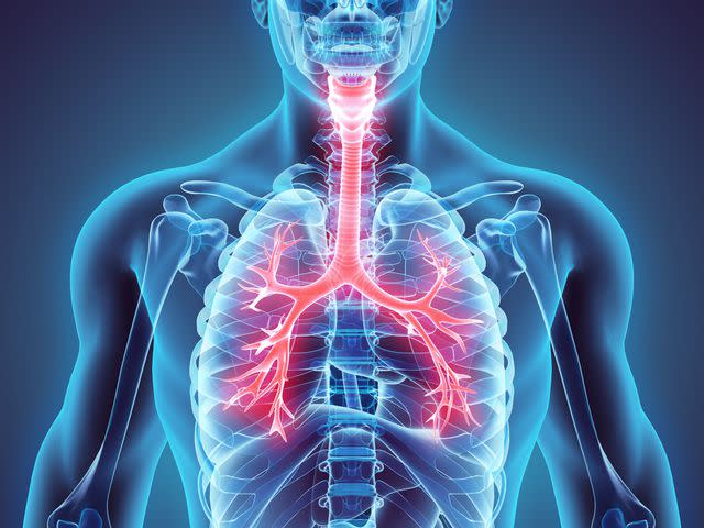
Tracheal stenosis is a narrowing of your trachea, or windpipe, due to the formation of scar tissue or malformation of the cartilage in the trachea. While mild narrowing in your trachea may never be identified, a significant narrowing of more than 50% of your airway can lead to serious complications. The ttwo most prevalent causes of tracheal stenosis are:
Prolonged placement of an endotracheal tube (breathing tube) or tracheostomy
Collagen vascular disease (granulomatosis with polyangiitis)
Other known causes include:
Congenital malformations (birth defect)
Trauma
Inhalation burns
Radiation therapy
Infections of the trachea
Inflammatory diseases (sarcoidosis or amyloidosis)
Cancer
In cancer and congenital malformations, the airway is being compressed either from outside the trachea or from narrowing from malformed cartilage.
Other causes of tracheal stenosis usually start with an ulceration in the trachea. The ulceration starts a cascade of inflammation, which is a normal healing process that can become exaggerated and may cause more scar tissue than would normally be necessary. This additional scar tissue narrows the area in your trachea.
Incidence
The frequency of acquiring tracheal stenosis depends upon the cause of the tracheal narrowing. Post-intubation damage to the airway can be common; however, the risk of symptomatic stenosis is low. The following risk factors will increase your likelihood of having post-intubation or tracheostomy-related tracheal stenosis:
Tracheal stenosis may be one of the first signs seen in granulomatosis with polyangiitis. Stenosis can occur about 20% of the time. There is not much data available on the prevalence of other causes of tracheal stenosis.
Symptoms
In congenital tracheal stenosis, mild stenosis can often be misinterpreted as asthma or recurrent bronchitis. With mild tracheal stenosis, you may not identify symptoms until later childhood or early adolescence when symptoms appear as difficulty breathing with exercise. In more severe cases of congenital tracheal stenosis, you may notice the following symptoms:
Stridor (high pitched breathing sound)
Cyanotic, with noticeably blue lips
Wheeze with inhalation
Exertional shortness of breath (dyspnea)
In other cases of acquired tracheal stenosis, the symptoms may not present themselves for several weeks after the injury occurs. Difficulty with breathing is the common first symptom. Like congenital tracheal stenosis, you may notice stridor, wheezing, or exertional shortness of breath.
Diagnosis
Several testing methods may be used to help your healthcare provider determine whether you have tracheal stenosis or not. Bronchoscopy is considered the gold standard for diagnosing tracheal stenosis because your healthcare provider will be able to directly visualize your trachea.
However, there are some risks associated with this because using a scope will further obstruct your airway, so maintaining your oxygenation levels may be more difficult. Discuss your individualized risk factors associated with bronchoscopy with your healthcare provider.
Other methods that your healthcare provider may use include X-ray, CT scan, ultrasound, MRI, and pulmonary function testing. Standard X-rays are good at the identification of structure, columns of air, trauma, and other preliminary data. Other more sophisticated X-ray machines can be used (xeroradiography) to further identify stenosis; however, the radiation exposure is significantly higher than other methods.
Computed Tomography (CT) Scans
CT scanning can be a great technique for your healthcare provider in determining whether you have tracheal stenosis or not. It does, however, have difficulty identifying soft tissue causes of the narrowing of your trachea. Some techniques are being utilized in a way to create "virtual endoscopy" to minimize the need for you to undergo a bronchoscopy.
Ultrasound
Ultrasound can be helpful in identifying the amount of air space in the trachea. This allows your healthcare provider to determine whether or not more testing may be necessary; however, due to the amount of cartilage around the trachea, accuracy of the test can be questioned because of shadowing effects caused by the reflection of the sound waves off the cartilage. Leave this test only to those highly skilled at identifying tracheal stenosis by ultrasound.
Magnetic Resonance Imaging (MRI) Scans
MRI scanning is also a great alternative method to help in diagnosing tracheal stenosis, and in children, it is being considered to become a standard method. The major drawback of MRI is the length of time you need to commit to have the procedure done and the blurring that can occur from normal breathing during the exam. Improved techniques are continuously being developed to improve the utilization of this technique in diagnosing tracheal stenosis.
Pulmonary Function Testing (PFT)
Pulmonary function testing can be performed in some healthcare providers' offices, or if unavailable, you will be sent to a pulmonary lab. This test can be used to determine how much of an impact the stenosis is having obstructing your breathing. This will be helpful in discussions regarding treatment options with your practitioner.
Treatment
Several options exist for treating tracheal stenosis, and several types of healthcare providers are trained in performing these procedures. Dilitations may be performed by a thoracic surgeon, an otolaryngologist (head and neck surgeon), or even some pulmonologists. Whichever type of practitioner you choose, be sure to discuss which options are the least invasive and have the potential for the best result for your individualized care.
Most treatments are endoscopic procedures requiring actual visualization of your trachea. If the area of stenosis is small, placing a stent, dilating your trachea with a balloon, or removing some of the scar tissue with a laser will help to minimize the stenosis.
During these procedures, your healthcare provider may also inject the tissue in your trachea with steroids to help minimize any swelling. Although often initially successful, there can be a high rate of recurrence with some of these procedures.
Tracheal Resection
For more severe tracheal stenosis, your healthcare provider may recommend tracheal resection, which requires surgery. This is a difficult operation and should be done by someone who does a lot of them to predict the best outcomes.
This procedure is reserved for when endoscopic treatments have failed, or tracheal stenosis is too severe for endoscopic procedures. During this procedure, your healthcare provider will cut out the part of the trachea that is affected and repair your trachea with skin or cheek tissue.
Follow-Up
Following surgery, you will typically be able to have the breathing tube removed during recovery from anesthesia. However, if there is too much swelling, several interventions will be used. In this case, you can expect to be placed on steroids, as well as a diuretic. Healthcare providers will also be sure to keep the head of your bed elevated. Shortly thereafter, you will return to the operating room to have your breathing tube removed. If you are still unable to support your airway, a tracheostomy will be inserted to maintain your airway. Due to the invasive nature of this treatment, it is considered a last resort after other therapies have failed.
Read the original article on Verywell Health.

