Stages of Macular Degeneration
Dry MD occurs in stages, while wet MD is typically diagnosed at an advanced stage
Medically reviewed by Andrew Greenberg, MD
Macular degeneration is an eye disease that affects your central vision. It is a common problem that ranks as the leading cause of vision loss among adults who are more than 60 years old. Because it is linked with aging, it is also called age-related macular degeneration (AMD).
Macular degeneration occurs due to the damage that aging causes to your macula. This part of your eye helps you see the details of what is in front of you. It is a small place at the center of your retina (the light-sensitive tissue at the back of your eye).
The causes of macular degeneration are not fully known. Symptoms can affect your central vision, though they do not cause total blindness. There is no cure. Treatments may help preserve some central vision based on the type and stage of your disease.
This article describes the stages of macular degeneration and their common symptoms. It also explains the treatments used at each stage and what they do.
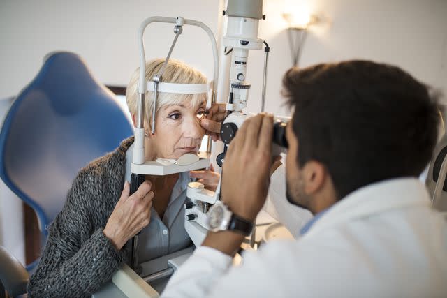
Martinns / Getty Images
Stages of Macular Degeneration
Macular degeneration is categorized into:
Early
Intermediate
Late stage
The late stage of the disease can occur as one of two macular degeneration types, as follows:
The dry form of macular degeneration is the most common type. It affects up to 90% of people with AMD.
Below are the stages in detail.
Early-Stage Dry AMD
Early-stage dry AMD usually occurs without noticeable symptoms. It results when parts of your macula thin with age and allow for the formation of drusen. Drusen are small clumps of lipid and protein that build up on the tissue of your retina.
Drusen can be identified during a routine dilated eye exam. Having a few small drusen can occur in people 50 and older without macular degeneration or vision loss. However, the presence of many small drusen or medium-sized drusen can often indicate early-stage AMD.
In some cases, early-stage dry AMD may be accompanied by a loss of dark adaptation, which is the ability of your eyes to adjust from a bright environment to a darker one. While this problem is common with age, it can happen earlier and at an accelerated rate with AMD.
Intermediate-Stage Dry AMD
When dry AMD progresses to the intermediate stage, large drusen and/or abnormalities in your retinal pigment are detected. While this stage may still be asymptomatic for some people, intermediate-stage AMD may involve one or more of the following more subtle symptoms:
Area of blurred or fuzzy central vision
Increased difficulty seeing in low-light conditions
Increased loss of dark adaptation
Loss of color vibrancy so that it is hard to tell the difference between similar colors or shades of the same color
The rate at which these changes occur can vary by individual. While some people may have more pronounced symptoms at the intermediate stage, the impact on vision at this stage may be minimal for others. It is also possible to have AMD without the disease progressing to late AMD.
Advanced or Late-Stage Dry AMD (Geographic Atrophy)
Advanced-stage dry AMD, also called geographic atrophy, accounts for 20% of all legal blindness caused by AMD. Research indicates that about 70% of its onset is linked to genetics while aging and factors like smoking, poor diet, and sun exposure contribute to 30%.
It occurs when dry AMD progresses to the point that clearly defined areas of your retina lose pigment and photoreceptors, causing areas of tissue atrophy (cell degeneration). This can result in irreversible vision loss. Up to 19% of people with AMD may progress to geographic atrophy within two years of diagnosis of intermediate dry AMD. This can occur with or without wet AMD.
Late-stage dry AMD results in the worsening of symptoms that occur during intermediate-stage dry AMD. This can lead to the following symptoms:
Blurry distance and/or reading vision
Hazy vision
Larger blank or blurry spot in your central vision, making it harder to read or recognize faces
Objects appearing smaller than their actual size
A blurred, gray, or empty spot in your central vision that interferes with reading and recognizing faces
Inability to recognize people's faces
Advanced Stage Wet-AMD
All AMD begins in the dry form. The condition changes to wet AMD in 10% to 15% of people. This can occur at any stage of dry AMD. There are no symptoms linked with early or intermediate stages of wet AMD. Wet AMD is only identified as an advanced-stage disease when it is diagnosed.
Though less common than advanced dry AMD, advanced wet AMD is a more serious condition. It occurs when new, abnormal blood vessels form under your retina. Your macula can become damaged when these new blood vessels leak blood and other fluids.
Symptoms of wet AMD are similar to those that occur at the intermediate and advanced stages of dry AMD. However, vision loss occurs much faster and more dramatically with wet AMD than dry AMD.
In addition to the symptoms mentioned with dry AMD, wet AMD may also be accompanied by the following vision problems:
Straight lines appearing wavy or crooked
A blurry area near your center of vision that increases or forms into blank spots
Advanced wet AMD differs from dry AMD in the following two key ways:
Vision loss occurs much faster and more dramatically with wet AMD than with dry AMD. Dry AMD can exist for years before it causes symptoms that interfere with activities such as reading and driving.
Wet AMD causes severe vision changes and vision loss immediately without warning. This differs from the gradual onset that typically occurs with dry AMD.
Stargardt's Disease
Macular degeneration can happen at any age. Stargardt disease is a common form of juvenile macular degeneration. It typically affects both eyes, causing a slow loss of central vision between age 5 and early adulthood. Stargardt disease is linked to genetic causes.
While there is no cure, treatments and assistive devices can help make the best use of a person's remaining peripheral vision, which is the vision you see out of the corners of your eye).
AMD Stages in Different Eyes
AMD can occur in one or both eyes. Research indicates that 64.5% of people with AMD have the same stage of AMD in both eyes simultaneously.
When AMD exists at different stages in each eye, your healthier eye may compensate for the vision loss caused in the eye with the more advanced disease stage. This can delay your recognition of symptoms because you may not notice changes in your vision.
When AMD is diagnosed in only one eye, your healthier eye is likely to develop AMD. Research indicates that having any stage of AMD in one eye is expected to affect the second eye within five years in 19% to 28% of people. When late AMD is diagnosed in one eye, the condition is likely to affect the second eye within five years in 50% of affected people.
The dry and wet forms of AMD can occur at the same time. The dry form may continue to progress in an eye that has developed the wet form. You can have dry AMD, wet AMD, or both simultaneously in one or both eyes.
Learn More: Macular Degeneration: Timeline of Vision Loss Progression
How to Get Macular Degeneration Staged
If you are at risk of AMD, monitoring your eye health with regular comprehensive eye exams from an ophthalmologist or optometrist is important. An eye exam includes a series of tests that measure your eye health and vision.
Age is the major risk factor for macular degeneration. Being age 55 or older increases your risk of AMD. The American Academy of Ophthalmology (AAO) advises more frequent eye exams as you age, based on the following schedule:
A baseline comprehensive eye evaluation at age 40
Every two to four years for people between the ages of 40 to 54 years who do not have symptoms or risk factors for eye disease
Every one to three years for people between the ages of 55 and 64 years
Every one or two years for people 65 and older, even if there are no symptoms or changes in vision
You may need eye exams more often based on factors such as your demographic characteristics, medical history, personal history of smoking, or a family history of eye disease.
The diagnosis and staging of macular degeneration typically involves one or more of the following tests:
Dilated eye exam: A dilated eye exam can help your provider identify a drusen (small piece of cell waste on your retina) or changes to your macula, which may be early signs of macular degeneration.
Visual acuity exam: Standard vision screening tests use the letter chart to measure your vision. An Amsler grid may also be used. This chart, which looks like a piece of graph paper, is used to determine whether you have blurry or blank spots in your field of vision.
Fluorescein angiography: This test involves the injection of fluorescein (a yellow dye) into a vein in your arm or hand that travels through your bloodstream. A special camera takes photos of your retina. If fluorescent patches appear, they may indicate leaking blood vessels in your retina (wet AMD).
Optical coherence tomography (OCT) and optical coherence tomography angiography (OCTA): OCT is a noninvasive imaging procedure that makes cross-sectional photos of your retina. It provides very detailed information about retinal structure, drusen, new blood vessels, and hemorrhaging. OCTA uses the same procedure to take pictures of the blood vessels in and under your retina.
Fundus autofluorescence imaging (AF): AF is another noninvasive way to examine the blood vessels under your retina. Instead of a fluorescent dye, AF uses the fluorescence of certain structures in your eye to study your retina. It can be useful in studying late dry AMD because the areas damaged by late dry AMD do not light up.
Macular Degeneration Staging vs. Progression
Disease staging is a system of classifying disease severity based on diagnostic findings. The stage of disease is used to determine your treatment and prognosis.
Disease progression describes the natural way that a disease changes, worsens, and possibly improves over time.
Treatment for Different Stages of Macular Degeneration
Treatments do not cure macular degeneration at any stage. However, appropriate treatments administered as early as possible can help slow the disease progression of AMD.
Early-Stage Dry AMD
Treatment of early-stage dry AMD focuses on identifying modifiable lifestyle risk factors and the actions you can take to slow the disease's progression. These actions can include the following:
Eat a nutritious diet rich in fruits, nuts, vegetables, dark leafy greens, and fish rich in omega-3 fatty acids.
Use an Amsler grid to perform self-screenings at home to track the progress of your disease.
Manage chronic conditions such as hypertension, obesity, and atherosclerosis.
Avoid-long-term exposure to bright sunlight without eye protection.
Continue with annual eye examinations unless symptoms suggest a change in disease progression and the need for an immediate examination.
Follow up with genetic testing if advised based on a family history of AMD.
Intermediate-Stage Dry AMD
A diagnosis of intermediate-stage dry AMD increases your risk of developing late-stage AMD and the vision loss that it can cause. Research from the Age-Related Eye Disease Study (AREDS) and AREDS2 studies signifies that certain antioxidants, vitamins, and minerals can help protect against the progression from intermediate to late-stage AMD.
The current recommended regimen of supplements, called AREDS2 supplements, can be purchased over the counter in a single formulation. They are taken daily and consist of the following ingredients:
Vitamin C: 500 milligrams (mg)
Vitamin E: 400 international units (IU)
Copper (cupric oxide): 2 mg
Zinc: 80 mg
Lutein: 10 mg
Zeaxanthin: 2 mg
Check with your ophthalmologist before taking the AREDS2 supplements because they may not be effective for treating all forms of the disease.
Recommendations for managing intermediate AMD and reducing your risk of disease progression also include the following:
Quitting smoking
Continuing self-assessment using an Amsler grid at home to track changes in your disease
Getting regular eye exams every six months or less, based on the schedule advised
Scheduling an urgent consultation with your ophthalmologist at the first sign of a sudden change in vision
Advanced or Late-Stage Dry AMD (Geographic Atrophy)
In 2023, the Food and Drug Administration (FDA) approved the first treatments for adults with geographic atrophy caused by advanced-stage dry AMD. These drugs include the following:
Syfovre (pegcetacoplan)
Izervay (avacincaptad pegol)
These in-office treatments are administered as intravitreal injections in the vitreous cavity (the space at the back of your eye behind the lens). The medications are injected into your eye on a scheduled basis to slow the atrophy, hoping to delay vision loss.
Advanced or Late-Stage Wet AMD
Treatment for late-stage wet AMD is used to reduce the number of abnormal blood vessels in your retina and slow any leaking from the vessels:
Anti-VEGF (anti-vascular endothelial growth factor) treatment: Anti-VEGF treatment is considered the primary treatment for wet AMD. The medication is administered every one to three months via an intravitreal injection. Current anti-VEGF therapies include the following medications:
Avastin (bevacizumab)
Lucentis (ranibizumab)
Vabysmo (faricimab-svoa)
Photodynamic therapy (PDT): Photodynamic therapy may be used with anti-VEGF injections. It works by injecting a light-sensitive medicine called verteporfin into your arm.
The medicine travels to your eye, where it is activated when lit by a certain type of laser. Shining the laser on a small area in the back of your eye triggers the verteporfin to break down the new blood vessels, causing vision loss.
Summary
Macular degeneration is an eye disease that occurs as a result of damage to your macula caused by aging. While the exact causes of the disease are not fully known, age is the main risk factor.
Stages of macular degeneration include early, intermediate, and late stages. Late-stage disease can involve wet or dry AMD. While there is no cure for the disease, treatments can help slow progression. They can also help prevent further vision loss in certain cases.
Always report any vision changes to your eye care provider even if you are not due for an exam. Some changes that occur with this disease can progress very quickly but may respond to treatment if given quickly.
Read the original article on Verywell Health.

