Retinoschisis: Overview and More
Medically reviewed by Bryan M. Wolynski, OD
Retinoschisis is a condition that causes splitting of the layers of the retina—the light-sensitive tissue in the back of the eye. The retina communicates with the brain about what the eyes see.
Having retinoschisis can affect a person’s vision; however, some people with the condition do not notice any changes to their eyesight. Retinoschisis usually affects both eyes, but it can also occur in each eye to different degrees.
Related: Retina: Anatomy and Function
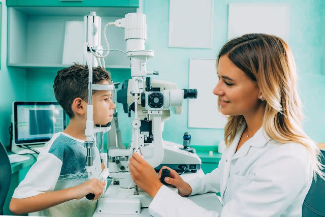
Stevica Mrdja / EyeEm / Getty Images
Types of Retinoschisis
There are two types of retinoschisis—one that a person is born with and one that develops as a person gets older.
Juvenile X-Linked Retinoschisis
This form of retinoschisis is a genetic disease that mostly occurs in young males. Overall, it affects one in 5,000 to one in 25,000 people.
Juvenile X-linked retinoschisis occurs when an abnormal gene is carried on the X chromosome. Biological males only have one X chromosome. If the abnormal gene linked to retinoschisis is on that one chromosome, a person can develop the condition and its associated vision problems.
Takeaway
Biological females can also have juvenile X-linked retinoschisis caused by an abnormal gene on the X chromosome. However, because they have a second X chromosome that is not affected, they usually have normal vision.
Degenerative Retinoschisis
Also called acquired or senile retinoschisis, this form is more common than the hereditary form. It occurs equally in males and females. Overall, it affects about 4% of people age 40 and older.
Retinoschisis With Cysts
In some cases of retinoschisis, small cysts grow on the retina and may damage the nerves, leading to changes in vision. While it is not common to go blind from retinoschisis, the form that is inherited can cause poor vision.
Related: How Genetic Disorders Are Inherited
Retinoschisis Symptoms
Retinoschisis does not always have symptoms, particularly with the degenerative form. More often, the condition is spotted during an eye exam. In children, vision changes may not be detected until they have a vision screening.
If a person does have symptoms of retinoschisis, they can include:
Blurry vision
Changes to vision that get worse with time
Darker, dimmer vision
Trouble seeing things from the side (peripheral vision)
People with the genetic form of the condition often experience abnormal eye movement and a clouding of the lens of the eye (cataract). They may also have other signs and symptoms, such as farsightedness and glaucoma.
When to See a Doctor
Vision changes and blurry vision can have many causes and are not always caused by retinoschisis. If you experience changes to your vision, an eye exam can help determine the cause.
If you develop sudden, new changes to your vision or you start to see flashes or small, floating objects in your vision, make an appointment with your eye doctor right away, as these can be signs of a more serious eye condition.
Causes
It’s not clear why some people get retinoschisis and others do not. However, it is known that the hereditary form is caused by an abnormal gene (mutation) and the degenerative form is associated with aging.
Related: Common Causes of Vision Loss
Diagnosis
Before diagnosing retinoschisis, eye doctors need to rule out similar-looking conditions, such as a detached retina or central serous chorioretinopathy. Retinoschisis also can be confused with conditions like “a lazy eye” (amblyopia).
The diagnosis and management of retinoschisis are usually handled by a type of eye doctor called a retinal specialist.
There are two main tests that eye doctors use to diagnose retinoschisis: electroretinograms and optical coherence tomography.
Electroretinogram
An electroretinogram test measures the electrical sensitivity of the retinal cells. During the test, an electrical sensor is placed on the eye to measure the retina’s electrical activity in response to light. The results are shown on a screen for an eye doctor to review.
Takeaway
Electroretinograms can be hard to do on children who are between the ages of 2 and 5. Kids in this age group may need to be put under general anesthesia to have the test. Some eye doctors only do the test on children younger than 2 or older than 5.
Optical Coherence Tomography
Eye doctors use optical coherence tomography to detect many conditions and diseases. The test uses light to make a high-resolution 3D image of the eye, especially the back portion.
The test is similar to an ultrasound, but instead of measuring sound, it measures light. The images are incredibly detailed and allow eye doctors to see things that they wouldn’t be able to see with other imaging scans like MRIs.
Related: Optical Coherence Tomography: What to Expect
Other Tests
There are also a few other ways that eye doctors can look for retinoschisis, including:
Measuring the eye’s visual evoked response to light
Ultrasonography or ultrasound
Genetic Testing
To diagnose the hereditary form of retinoschisis, eye doctors might use genetic testing. While females can be carriers of retinoschisis, they do not usually have findings associated with the condition on tests.
A family tree analysis can help eye doctors explain to patients how the condition might be passed on—for example, juvenile X-linked retinoschisis regularly affects male family members.
Genetic counseling can be helpful for people with the genes associated with retinoschisis.
Complications
Prompt diagnosis and management of retinoschisis are essential to prevent complications related to the condition, which can threaten a person’s vision.
Retinal Detachment
Having retinoschisis raises a person’s risk for developing a potentially vision-threatening condition called retinal detachment—when the retina separates from the back of the eye. Retinal detachment occurs in 5% to 22% of people with the inherited form of retinoschisis.
If a detached retina is found early, eye doctors can treat it with surgery to help prevent permanent vision loss.
Related: What Is Retinal Detachment?
Bleeding
Another complication that can occur with retinoschisis is bleeding of the gel that fills the back of the eye. The gel is called the vitreous; when it bleeds, it’s called a vitreous hemorrhage.
If a person develops a vitreous hemorrhage, an eye doctor will use laser or cryotherapy to close up the damaged area of the retina that is causing bleeding.
Takeaway
People with retinoschisis are sometimes advised not to take part in high-contact sports or activities that raise the risk for a retinal detachment or vitreous hemorrhage.
Cysts
Sometimes, a type of eye medication called carbonic anhydrase inhibitors can help treat the cyst spaces that occur during X-linked retinoschisis.
Treatment
People with juvenile X-linked retinoschisis need regular eye exams—every six months to a year—to monitor the condition’s progression.
People with the degenerative form of the condition often do not need specific treatment. However, if they have symptoms or certain risk factors for complications, they do have options for managing the condition.
Related: Conditions Eye Exams Can Detect
Vision Aids
When retinoschisis impairs a person’s vision, low vision aids can help them see better during their day-to-day activities. Glasses can also help improve vision, but they will not fix the retina damage caused by retinoschisis.
Surgery
People with degenerative retinoschisis usually do not need treatment. However, if they develop a complication like retinal detachment, surgery might be recommended.
Related: Surgery for Retinal Detachment
Prognosis
The outlook for a person diagnosed with retinoschisis depends on whether they have the genetic form of the disease or the type that develops with age.
The inherited form of retinoschisis is a lifelong disease that requires regular eye exams and careful monitoring. The degenerative form that can occur with aging usually does not progress.
Coping
If you or a loved one is diagnosed with retinoschisis, keeping up with your eye doctor appointments will be important to your eye health.
If you have the genetic form, you will likely need to work closely with your eye doctor to monitor the condition’s progression and protect your vision.
If you have the form that develops as you get older, follow up with an eye doctor or retinal specialist as indicated by them.
Related: Helping a Loved One Cope With Vision Loss
A Word From Verywell
Retinoschisis is an eye condition that causes splitting of the layers of the light-sensitive tissue in the back of the eye (retina). It can affect one or both eyes and can be inherited or acquired.
The genetic form of the condition typically occurs in young males and requires lifelong monitoring to prevent complications. The other form, which is more common, develops as people get older. It usually does not progress or require specific treatment; however, people who have vision problems related to the condition might benefit from using low-vision aids to help them with their activities of daily living.
Having retinoschisis can increase a person’s risk of vision-threatening complications like retinal detachment. Regular eye exams can detect these problems early and help eye doctors treat them early enough to protect a person’s vision.
Read the original article on Verywell Health.

