Researchers bioengineer skin, bioprint cartilage to fix ear malformation
Researchers in Switzerland and the United States have combined bioengineered human skin with bioprinted cartilage to produce what in the future could be perfect-looking ears to replace the outer shell of an ear that developed wrong while a baby was in the womb.
It’s among the latest advances in bioengineering and bioprinting. Earlier this month, Wake Forest researchers created full-thickness human bioprinted skin that may one day be an essential part of wound healing and scar reduction.
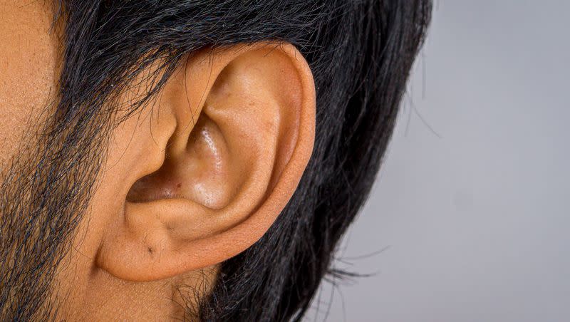
The congenital ear malformation is called microtia, which in Latin means “little ear.” And the study, published in the journal Science Advances, notes that the malformation of the external ear can create psychosocial problems for the child.
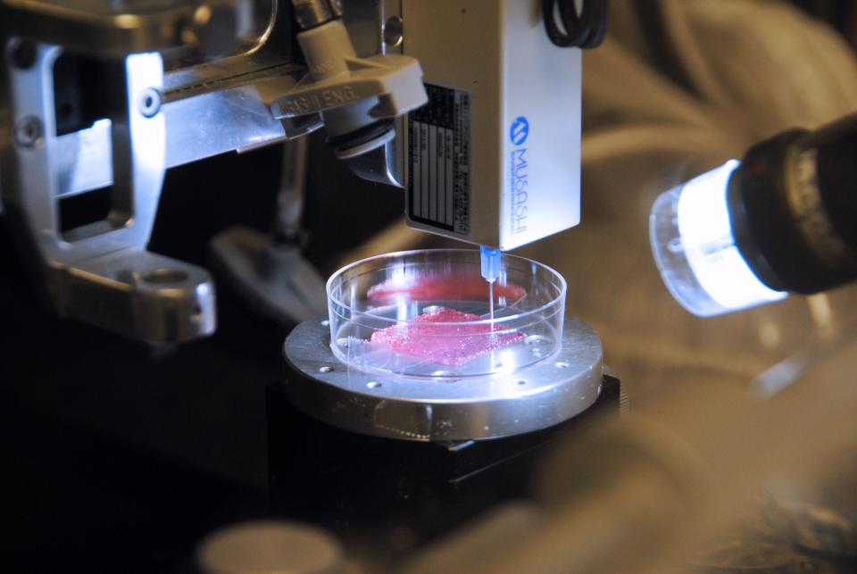
The researchers call the bioprinted cartilage EarCartilage and the “bioengineered human-pigmented and pre-vascularized” skin substitute EarSkin. Results from testing in rats have been promising.
The bioengineered ears have been created to include blood capillaries that in studies in immunocompromised rats connect to the recipient’s blood vessels within a week. And the bioengineered EarSkin has cells that contain pigments that “efficiently restored the skin color.” So the two together “represent a novel approach for the treatment of microtia with the potential to circumvent existing limitations and improve the aesthetic outcome” of outer ear reconstruction, the researchers write.
Existing remedies for microtia
The bioengineered result is different from a prosthetic ear, which is made to look like an outer ear and simply attaches. Silicone is commonly used as the material in ear prosthetics.
A number of more natural options have in the past been used to treat microtia using the patient’s existing outer ear, as long as the microtia is mild and the outer ear relatively well-formed. The Microtia and Atresia Center at Vanderbilt has had great success with pre-formed ear molds or ear expanders soon after birth in cases where the ear is well-formed but small. Slight malformations have also been treated that way. They also have other nonsurgical options for slight malformations in very young babies, taking advantage of the fact that ear cartilage is soft in infants.
Medicalxpress.com notes the “gold standard” treatment of microtia has involved taking cartilage from the rib cage and creating a graft framework. “This construct is then implanted under the skin of the skull to elevate the auricle in a second stage in a highly complex plastic surgery procedure. The intervention required harvesting a sufficient amount of costal cartilage and is therefore usually delayed until children are 10 years of age.“
The article says of the Zurich team’s new effort: “The approach presented an innovative and promising treatment strategy for patients born with microtia,” overcoming some of the challenges encountered with earlier efforts.
Printing skin that heals
Bioengineering is also being used to tackle other tissue and skin needs.
The Wake Forest Institute for Regenerative Medicine this month published a research paper in Science Translational Medicine on a “significant breakthrough in the area of skin regeneration and wound healing,” according to the university’s press release.
“Skin regeneration has long been studied with hopes of providing burn victims, wounded warriors and those with skin disorders opportunities at complete healing. Available grafts are often temporary, or if permanent, have only some of the elements of normal skin, which often have a scarred appearance. The creation of full-thickness skin has not been possible to date,” the university said.
The researchers bioprinted all six major human cell types that are normally found in skin, combined with “specialized hydrogels as a bioink.” The resulting full-thickness skin included the three layers found in normal human tissue: the epidermis, dermis and hypodermis.
The researchers said that when transplanted in early studies, the bioprinted skin “formed blood vessels, skin patterns and normal tissue formation.” In further studies, they showed the bioengineered skin improved wound closure and helped reduce scarring.
“Comprehensive skin healing is a significant clinical challenge, affecting millions of individuals worldwide, with limited options,” said study co-leader Dr. Anthony Atala, primary author on the paper. “These results show that the creation of full-thickness human bioengineered skin is possible, and promotes quicker healing and more naturally appearing outcomes.”

