Psychedelic look into a rat's eye wins microphotography competition
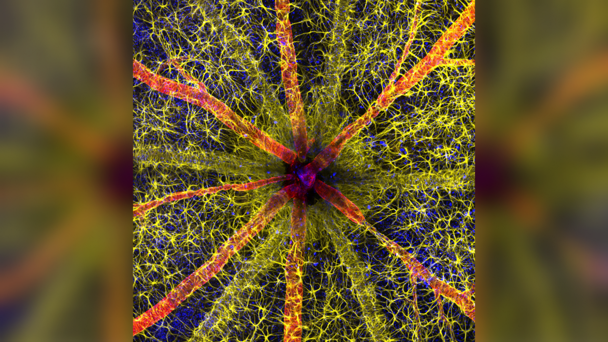
A psychedelic image of the spider web-like network of tiny vessels in the back of a rat's eye has won this year's Nikon's Small World photography competition.
Hassanain Qambari, a diabetes researcher at the Lions Eye Institute in Australia, was awarded first prize for the detailed image of a rodent's optic nerve head. Along with Jayden Dickson, Qambari submitted the image to the competition "to showcase the complexity of retinal microcirculation," Qambari said in a statement.
The researchers study diabetic retinopathy, a condition in which long-term high blood sugar levels damage the small vessels of the eye, potentially causing complete vision loss.
"Current diagnostic criteria and treatment regimens for diabetic retinopathy are limited to the late-stage appearance of the disease, with irreversible damage to retinal microvasculature and function," Qambari said.
One of the challenges they faced was locating these tiny vessels, which are around 0.004 inch (110 micrometers) in diameter. To create the intricate image, Qambari collected and stacked hundreds of single images taken with confocal and fluorescence microscopy.
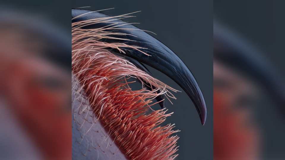
Photographer John-Oliver Dum, with the company Medienbunker Produktion in Germany, was awarded fourth place for his terrifyingly beautiful shot of venomous tarantula fangs. In the haunting close-up, the large, rounded black fangs of a Caribena versicolor glow red at the tips and are surrounded by a dense collection of pink hairs. The picture was taken in collaboration with the Museum of Natural History in Chemnitz, Germany.
"We had planned that I would give pictures for a lecture about the microworld of their insects and spiders, to show the unknown world," Dum told Live Science in an email. But due to the COVID-19 pandemic, this project was never finished.
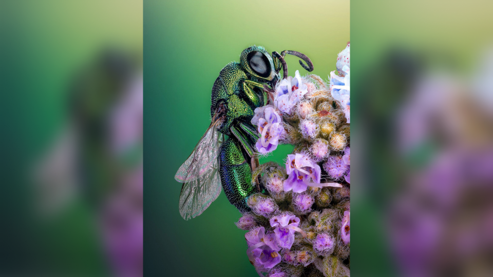
Photographer Sherif Abdallah Ahmed, from Tanta University in Egypt, was awarded 12th place for a stunning photo of an iridescent, emerald-green cuckoo wasp (Chrysididae) perched on a mass of purple flowers. Much like cuckoo birds, cuckoo wasps are kleptoparasites — they lay their eggs in other wasps' nests. The larvae of the thieving wasps then devour all the food stored in the nest, sometimes along with any other eggs there.

Yuan Ji, from the World Expo Museum in Shanghai, was awarded 17th place for a colorful, ethereal photo of a Chinese moon moth's (Actias ningpoana) wing scales. These moths live in the mountainous cloud forests in China. Bats hunt these moths, but the moths use their twisted tails to sabotage the bats' echolocation, lowering the risk of being caught.
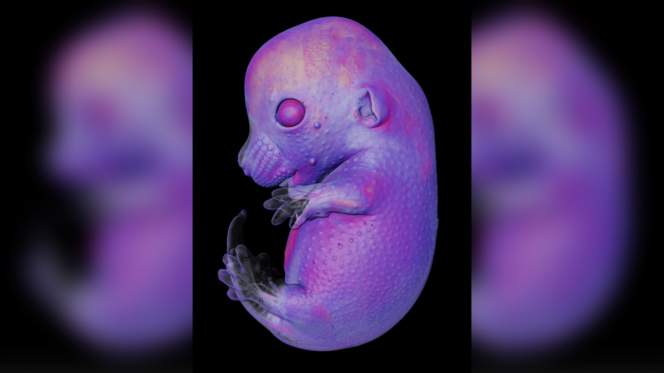
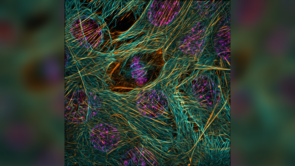
Other noteworthy images included a colorful view of defensive hairs covering the surface of a leaf, a glimpse into the development of a mouse embryo and an intricate view of cell division in a myoblast stem cell.
RELATED STORIES
—Glowing embryonic gecko hand and otherworldly slime mold amaze in winning microscope photos
— Electrifying time-lapse video shows neurons shooting across the inside of a chick embryo
— 'Hauntingly beautiful' image of a golden horseshoe crab wins wildlife photography competition
Nikon's Small World photography competition showcases the beauty of scientific imagery captured through the lens of a light microscope. This year's winners were announced Oct.17.
Nikon's Small World competition also includes an award for videography. On Sep. 26, the Nikon Small World in Motion video competition top prize was awarded to Alexandre Dumoulin for his 48-hour time-lapse video of developing neurons in the central nervous system of a chick embryo.

