What Is the Palatine Bone?
This bone helps form the nasal cavity, eye socket, and palate
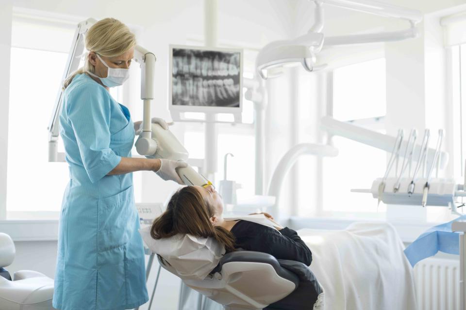
Portra / Getty Images
The palatine bone (os palatinum) is a paired, L-shaped facial bone that makes up a portion of the nasal cavity and palate. It lies between the maxilla bone (the fixed, upper bone of the jaw) and the sphenoid bone, the wings of which help form the base of the eye sockets and base of the skull.
Numbing done as part of molar and premolar tooth extractions can sometimes affect the nerves that run through the palatine bone, and the bone itself can be fractured in an accident. It may also be involved in a cleft palate diagnosis, typically at birth.
This article presents the anatomy, structure, and function of the palatine bone. It also discusses conditions that affect it, as well as how these conditions may be diagnosed and treated.
Anatomy of the Palatine Bone
The palatine bone is actually two bones that come together. They are L-shaped, mirror images of one another and join where their horizontal parts meet up. This is down the middle of the upper mouth at the median palatine suture.
On the upper border, the palatine helps form the base of the orbital process.
The location of the palatine bone is best understood through its borders and articulations, where it connects to other structures.
Horizontal Plate
The horizontal plate is composed of the two parts that make up the bottom of the L-shaped bones. It makes up the roof of the mouth and the rear portion of the oral cavity, just behind the nasal cavity. The front end is serrated and the back end is smoother.
More specifically, it sits just behind the maxilla bone of the upper jaw, lying in front of the soft palate (the soft tissue at the roof of the mouth).
The two palatine bones give rise to the posterior nasal spine towards the back of this plate. This part also includes the greater palatine foramen, a space that contains the greater palatine nerve as well as its necessary blood vessels.
Vertical Plates
The perpendicular plate is the vertical part of each palatine bone's "L" shape. It makes up a portion of the sidewall of the nasal cavity at the point where it joins the sphenoid bone and pterygoid process (projections from the sphenoid that are essential for jaw and mouth movement).
This plate also gives rise to the orbital process, which makes up a part of the socket where the eye sits.
Here, the greater palatine canal, which runs between the sidewall of the palatine bone and the adjacent maxilla bone, is also observed. This portion also includes a sphenopalatine notch on the upper border, which connects with the sphenoid bone.
Pyramidal Process
The last part of the palatine bone is a pyramid-shaped area called the pyramidal process. This is located at the juncture between the horizontal and perpendicular plates (the corner where the two lines that form the "L" meet).
The lesser palatine canals are found here. These are spaces that house a range of important nerves and arteries.
Related:The Anatomy of the Maxilla
Anatomical Variations
The most commonly seen anatomical variation in the palatine bone has to do with the positioning of the greater palatine foramen, an opening toward the rear side that allows the descending and greater palatine nerves to pass through.
One study found that in approximately 73% of cases, this opening was located opposite the third upper molar tooth. It also noted a positioning opposite the second molar about 7% of the time, and in between the second and third molar roughly 16% of the time.
While subtle, variations of the palatine bone have significant clinical implications, especially for dentists or dental specialists performing molar or premolar tooth extractions.
Function of the Palatine Bone
Primarily, the palatine bone serves a structural function, with its shape helping carve out important structures within the head and defining the lower wall of the inside of cranium.
This bone helps form the nasal and oral cavities, the roof of the mouth, and the lower portion of the eye sockets (orbits).
As noted above, they also have openings that allow the palatine nerves to pass through. In this sense, palatine bones help house primary pain-signaling pathways for the mouth and teeth.
Related:Anatomy of the Greater Petrosal Nerve
Associated Conditions
There are several conditions related to the palatine bone. Nerve damage and fractures are possible, as are congenital (from birth) deformity and bone outgrowths.
Nerve Damage
The palatine bone is most often considered in dentistry because of the greater and lesser palatine nerves, known to be extremely sensitive. When dentists need to extract the upper molars and premolars, these nerves have to be anesthetized (numbed).
Sites of injection need to be carefully monitored—they typically are about 1 centimeter (cm) from the gingival margin (the “height” of the gums)—as there’s a risk of the syringe penetrating the greater palatine foramen.
The same care needs to be taken in children and adolescents, as their nerve pathways continue to develop.
There are clinical guidelines in place to prevent such nerve damage. Dental professionals and specialists, in particular, need to be versed in the variant anatomy of this bone.
Related:Numb Face
Fractures
Palatine bones can fracture due to accidents or falls. These palatal fractures are relatively rare and occur most often in adult men. They present a difficult challenge for healthcare providers because of their position in the face.
There are six major palatine fracture types based on the location of the bone break:
Anterior and posterior alveolar (often caused by trauma to the face and teeth)
Sagittal
Para sagittal
Para alveolar
Complex
Transverse fractures, often accompanying Le Fort fracture of the maxillary bone in the jaw
Not only can surrounding structures be affected, leading to pain and swelling, but these issues can also lead to misalignment of the teeth and bite.
Palatal fractures are detected using medical imaging methods, usually computed tomography (CT) scans paired with X-ray. This allows doctors to assess the scope and location of the issue.
Related:Oral and Maxillofacial Surgery: Everything You Need to Know
Cleft Palate
Cleft palate is a congenital deformity typically diagnosed at birth. It happens when the two halves of the palatine bone don't joint together during fetal development. Cleft palate and cleft lip are relatively common, occurring in roughly one in 500 to 700 births.
People born with a cleft palate may have dental problems, as well as difficulty with speech, hearing, eating, and drinking. They might also have frequent colds and ear infections.
Related:Cleft Lip and Palate Types
Torus Palatinus
Torus palatinus describes the development of mostly benign, painless outgrowths from the palatine bone. These tend to arise in the mid-plate of the palate and can occur on one or both sides.
Though usually asymptomatic, and often never noticed by patients, some cases do lead to:
Pain
Ulcers in the mouth
Disrupted chewing
Impaired speech
Torus palatinus is a condition that arises most often in adults in their 30s.
Treatment
Treatment depends on the issue affecting the palatine bone.
Nerve Damage Treatment
For minor nerve damage, a healthcare provider may recommend waiting to see if the issue resolves itself. Most of these injuries get better on their own within six months.
If symptoms are prolonged or severe, treatment options may include:
Corticosteroids, which are anti-inflammatory medications
Surgery to re-connect the nerve or fill in a gap between the nerve (called a nerve graft)
Laser therapy
Fracture Treatment
Treatment varies based on the severity and location of the fracture. There are two surgeries that may be considered:
Open reduction and internal fixation (ORIF)
Intermaxillary fixation (IMF)
In both cases, the idea here is that surgeons access the fractured bone, correct any alignment problems, and use splints, orthodontic braces, arch bars, or other methods to set in place.
Pain and inflammation need to be managed following surgery, with the length of recovery depending on how severe the fracture is.
In recovery, which usually takes three to four weeks, pain and inflammation are managed with prescription drugs.
Cleft Palate Treatment
Treatment of a cleft palate almost always requires a surgery called a palatoplasty, which aims to close the opening between the nose and mouth.
Surgical repair to correct cleft palate is ideally done at about 9 months of age, although it may be done up to age 2. It is performed by a plastic surgeon in conjunction with a team of medical specialists, including dentists, otolaryngologists, and speech pathologists.
Children are then reassessed for speech development and other factors at school age.
Some cases of cleft palate have been repaired in adults. While cleft palate surgery is generally safe and successful, it is not uncommon for changes made during the procedure to have later impacts.
Torus Palatinus Treatment
In cases where torus palatinus becomes symptomatic, or if it disrupts chewing and speech ability, doctors employ surgery to alter the shape of the palatine bone and remove the growth. Typically, this involves an incision in the middle of the palate to allow surgeons to get at the problem.
Treatment may be recommended if there's a reason for it, such as if the growths interfere with chewing or dental work. Surgery is the only way to remove the growths.
Summary
The paired palatine bone is a key structure of the skull, face, nose, and orbitals (eye sockets) while connecting to other bones, such as the sphenoid. It provides protection and passage for important nerves and blood vessels.
Palatine nerves need to be numbed during certain tooth extractions. Palatine bones also can be fractured and require surgery. Among the most common conditions involving the palatine, though, is a cleft palate diagnosed at or before birth, with repair typically successful.
The palatine bone primarily serves a structural function. If you have concerning symptoms or suspect an injury, be sure to talk to your healthcare provider in order to get an accurate diagnosis.

