What Is a Chest X-Ray?
Also Called a Radiograph
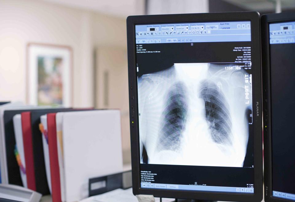
Reza Estakhrian / Getty Images
A chest X-ray is a medical test examining the inside of the chest. Also called a radiograph, this test gives the healthcare provider information on how well the lungs, heart, and other structures inside the chest are working.
This article will cover all the aspects of a chest X-ray, its risks, and how it works. It will also discuss what to expect during the test.
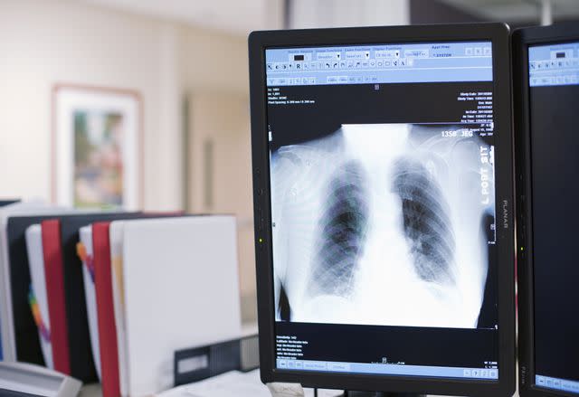
Reza Estakhrian / Getty Images
Purpose of a Chest X-Ray
A chest X-ray uses ionizing radiation to look at structures inside the chest. This test examines the lungs, heart, and chest wall to diagnose many medical conditions. A chest X-ray can also help healthcare providers monitor the progress of a medical condition to determine if treatment has been effective.
There are several medical conditions or reasons someone would need a chest X-ray. They may include:
Persistent cough
Enlarged heart
Chest pain
Chest injury
Broken bone
Cancer or tumors
Fluid in the lungs
A chest X-ray makes images of the inside of the chest. These images show the structures in black, white, and gray shades. This color variation is due to how much radiation the tissue absorbs. Bones absorb the most radiation and show up as white. Soft tissue and fat absorb less radiation and look gray. Air absorbs the least amount of radiation. Therefore, the lungs will look black.
X-rays are one of the most common diagnostic imaging tests because they are fast and require little preparation. A chest X-ray can provide healthcare providers with information on the structures inside the chest.
X-rays differ from a computed tomography (CT) scan or magnetic resonance imaging (MRI), which are also common imaging tests, in the following ways:
A CT also uses radiation to take pictures inside the body. However, a CT takes multiple images as it moves around the body. Then the computer combines all those images to create cross-sectional pictures, sometimes called slices. A CT uses more radiation than a chest X-ray.
An MRI produces a very detailed image of every structure in the body. It does not use radiation. While there are many benefits to an MRI, it does take much longer than a chest X-ray and is more costly.
A healthcare provider will choose a chest X-ray over a CT or MRI if they need fast imaging on a structure seen through an X-ray.
Risks and Contraindications
A chest X-ray does carry risks. Chest X-rays use ionizing radiation, which is a type of radiation that can cause DNA damage. The risks of ionizing radiation include:
These effects are typically seen at high levels of radiation exposure and are fairly rare.
While there are very real risks of radiation exposure during a chest X-ray, it's important to put the risk into perspective. Relative to most diagnostic imaging tests, a chest X-ray uses a small amount of radiation. One chest X-ray exposes you to 0.1 millisievert (mSv). People are exposed to 0.1 mSv over 10 days in their natural environment.
A healthcare provider must weigh the benefit vs. the risk of a chest X-ray. For instance, the test may be necessary to determine a medical condition or guide a healthcare provider's decision-making. If there is a medical need and no other diagnostic test can provide the same image quickly using less radiation, then the benefit generally outweighs the risk.
Notify a healthcare provider if you are or may be pregnant. Pregnant people can get a chest X-ray, but X-rays are avoided as much as possible to reduce radiation exposure to the fetus.
How to Prepare for a Chest X-Ray
When a healthcare provider recommends a chest X-ray, you should learn when and where to have it.
Sometimes chest X-rays are done during an emergency department (ED) visit and will happen right away. Other times, a healthcare provider may recommend one during a routine or sick visit. In these cases, the X-ray may not happen until later in the day and at a different location.
Timing
The actual imaging of a chest X-ray only takes seconds. However, it will take time to prepare for the chest X-ray. You may need to change clothes, walk to the radiology room, and get set up on the X-ray table. It is difficult to know exactly how much time this will take.
If the chest X-ray is performed during an emergency visit, the results are typically given during the stay. However, if the chest X-ray is done at an outpatient facility, the results are usually not given immediately.
Location
Chest X-rays can be performed in many locations. These include:
Outpatient healthcare facilities
Free-standing diagnostic imaging facilities
What to Wear
A chest X-ray needs a clear picture of the body. Most looser-fitting clothes are acceptable. You may be asked to change into a hospital gown. You won't be able to wear jewelry during the X-ray, so to be safe, leave it at home.
You also may be required to remove clothes that contain mental and cover the chest, like bras with underwires or shirts with metallic buttons.
Food and Drink
There usually are no restrictions on food and drink before a chest X-ray. However, follow any healthcare provider's instructions before the test.
Cost and Health Insurance
The cost and health insurance coverage will vary from provider to provider. A chest X-ray does not usually need preapproval, but checking your insurance plan for details is important.
What to Bring
When going to a chest X-ray appointment bring a health insurance card and identification.
The test will be short, and sedation is not used. You will be able to drive yourself to and from the appointment.
How Is a Chest X-Ray Performed
A chest X-ray is a relatively quick test that includes a few steps along the way. Below describes everything from the moment you enter the building until you leave.
Pre-Test
When you walk into the building for a chest X-ray, you must first check in. This is where insurance information and other basic information are gathered. If you do not have a history with the facility, they may have you fill out a health history form in the waiting room.
From the waiting room, you will be moved into a private area to change into a gown, if necessary. A radiology technician may ask you additional questions and explain how the test will proceed.
Throughout the Test
You will be moved into the room where the chest X-ray will be taken. A chest X-ray is typically done by taking two images. One will be taken from the back and one from the side.
For the image taken from the back, the X-ray technician will have you stand with your chest and hips pressed against the X-ray plate. For the side image, you will stand with your side against the plate and your arms elevated.
The X-ray technician may move into another room or behind a wall when in position. They will ask you to hold your breath briefly, and the X-ray will be taken. This should only take seconds.
Post-Test
Immediately after the test, the X-ray technician will ensure the images are clear and satisfactory for the radiologist to interpret. The radiologist is the healthcare provider who reads the X-ray and gives the results.
There is no recovery time or side effects after a chest X-ray.
Interpreting the Results
After a chest X-ray, a radiologist will review the images. They will provide their interpretation of the results and send them to your healthcare provider. Your healthcare provider will inform you of the results over the phone or may ask you to come into the office to discuss them.
When you are in the ED, the chest X-ray results will be given during that emergency visit.
Follow-Up
Depending on the results, follow-up may or may not be needed. If the radiologist is not able to visualize certain areas of the chest, they may need a repeat chest X-ray. Or if the chest X-ray does not provide the information necessary to make a diagnosis or guide the healthcare provider, then additional testing like a CT or MRI may be ordered.
Summary
A chest X-ray is a medical test that looks at the inside of the chest to provide healthcare providers with information about how well the heart, lungs, and other structures are doing. An X-ray does use radiation, but the amount is very small. There is almost no preparation for a chest X-ray, and the test is done within seconds.

