The Anatomy of the Sphenoid Bone
The sphenoid bone (wasp bone) is part of the base of the skull
Medically reviewed by Anita C. Chandrasekaran, MD, MPH
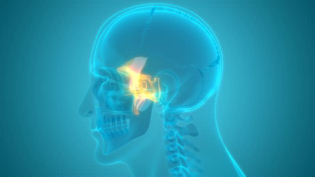
An unpaired bone located in the cranium (or skull), the sphenoid bone, also known as the “wasp bone,” is located in the middle and toward the front of the skull, in front of the occipital bone and behind the frontal bone.
The sphenoid bone is one of the seven bones that make up the orbit (the space that holds the eyeball), and helps make up the floor of the middle cranial fossa, the butterfly-shaped depression at the base of the skull that houses the temporal lobes of the cerebellum.
A central bone within the skull, it has a very complex shape, with a body and two sets of wings—the lesser and greater wings—as well as two pterygoid processes (protrusions that descend from where the wings meet the body).
Disorders or problems of the sphenoid bone can lead to a number of issues, including sphenoid sinusitis (an infection of the sphenoid sinuses), fractures, or sphenoid wing dysplasia—a malformation or deficiency due to a condition called neurofibromatosis type 1.
Anatomy
Structure
The sphenoid bone has a butterfly-like structure, with four major components—body, lesser wings, greater wings, and pterygoid processes.
Body
Located in a central portion that runs along the midline, the body of the sphenoid bone rests between the wings and forms several important structures. The front-facing portion helps make up the nasal cavity, while its sides contribute to the formation of the optic canal, a tunnel that allows the optic nerve and ophthalmic artery to pass through.
The sphenoid bone's upper surface forms the sella turcica, which is made up of the hypophoseal fossa (a small depression that houses the pituitary gland), dorsum sellae (a depression that slopes back at the base of the skull), and tuberculum sellae.
The sella turcica is surrounded by two anterior clinoid processes and two bony protrusions (one on each side), while at its rear are two other eminences called the posterior clinoid processes. These deepen the sella turcica and are attached to the tentorium cerebelli, a portion of the dura mater of the brain.
Lesser Wings
Rising from the front of the body of the sphenoid bone and moving off to the sides, the two paired lesser wings are triangular in shape. Their lower borders help make up part of the orbits (which house the eyes), while the upper surface makes up a portion of the cranial cavity, which houses parts of the brain.
Greater Wings
Emerging behind the lesser wings and also running to the sides are the two greater wings, which are also triangular and run lateral to the body. Their sides make up the infratemporal surfaces, which are convex in shape, and move backward and to the sides. These form parts of the infratemporal fossa, which are gaps at the base of the skull that allow nerves and blood vessels to pass through.
The front portions of the greater wings help form the sidewalls of the orbit. Each wing contains three openings—the foramen rotundum, foramen ovale, and foramen spinosum—which allow important nerves and vessels to pass through. The maxillary nerve, mandibular nerve, and middle meningeal vessels, respectively, pass through these openings.
Significantly, the superior orbital fissure, a large gap that allows nerves associated with vision to pass through, is at the border of the body and the lesser and greater wings.
Pterygoid Processes
Essentially extensions of the body of the sphenoid bone, the pterygoid processes consist of two protrusions emerging from the back of the body and continuing downward. These include two canals—the pterygoid and the palatovaginal canals, which allow nerves to pass through—and each has a hamulus, or a smaller, hook-like projection.
Location
The sphenoid bone rests in the central portion of the skull along its midline, separating the frontal bone (the forehead bone) from the occipital bone, a trapezoidal bone that makes up the lower back of the skull.
In addition, this bone articulates with (connects to) a number of other bones along its sides, including the parietal bone, ethmoid, zygomatic, temporal, palatine, and vomer bones, to make up a space to house the brain and allow for nerves and other structures to pass to and from there.
Takeaway
Largely, the sphenoid bone represents the floor of the skull.
Anatomical Variations
Given the complexity of this bone, it’s little wonder that there are a number of congenital variations in its anatomy. These can be divided into pneumatization (the presence of holes in the bones) and protrusion (extension of the bone), and include the following.
Pterygoid process pneumatization: In 15.5% to 43% of cases, healthcare providers have found pneumatization—that is, the presence of small holes—in the pterygoid process. This can occur on one side (unilaterally) or on both sides (bilaterally).
Anterior clinoid process pneumatization: The formation of holes has also been observed in the anterior clinoid process, which is located toward the back of the body of the sphenoid bone. As with the pterygoid process, this is seen either unilaterally or bilaterally, and has been found to occur in about 13% to 16% of people.
Foramen rotundum protrusion: Extensions of extraneous bone into the foramen rotundum have been reported in approximately 17.5% of cases. As with some other variations, this can occur unilaterally or bilaterally.
Internal carotid artery (ICA) protrusion: The ICA, a paired artery that runs up the sides of the neck and accesses the skull, has been observed to protrude into the sphenoid sinus and related areas. This has been reported in 12.75% of cases.
Pterygoid canal protrusion: Sometimes, small protrusions arise from the pterygoid processes into the pterygoid canal. This has been reported to occur in between 7.5% and 13% of people.
Function
Working in concert with the orbital floor, the primary function of the sphenoid bone is to help form the base and sides of the skull. Portions of this bone are also components of the facial skeleton.
Its central position within this part of the body makes it essential for providing rigidity—thereby protecting brain and nerve structures—while its rear parts are also attachment sites for muscles involved with chewing and talking.
Notably, too, the foramina (gaps) and the fissures of the sphenoid bone allow passage of important nerves and vessels in and out of the skull. Furthermore, a cavity in its body forms a sinus (called a sphenoid sinus) that connects to the nasal cavity; this allows the skull to be lighter and improves resonance.
Associated Conditions
There are several conditions that can affect the sphenoid bone; given its significance, these can have significant complications. Associated conditions include those below.
Sphenoid Sinusitis
The word "sinusitis" refers to an inflammation of the sinuses. It is often caused by a viral infection, but it can also be caused by bacteria or fungus.
Localized sphenoid sinus infection is rare but can be serious. The most common symptom is a headache, but other symptoms may include:
Ear pain
Pain behind the eyes
Pain around the temples
Takeaway
Most cases of sinusitis do not involve the sphenoid sinus. Typical viral sinusitis usually resolves in 1 to 2 weeks without treatment, but bacterial and fungal infections may need treatment with antimicrobial medication.
Symptoms of sinusitis include:
Sinus congestion or pressure, often localized to one area of the face
Headache
Post-nasal drip
Cough
Fever
Infection of the sphenoid sinuses can lead to either acute or chronic sphenoid sinusitis. Usually occurring alongside infection in surrounding areas, this condition can lead to fever, post-nasal drip, and weakness.
Seeking out prompt treatment is essential, since, if the infection is allowed to run its course, patients may develop severe issues, such as meningitis, brain abscess, and/or problems with cranial nerves.
Related: What Is a Sinus Infection?
Sphenoid Fractures
When fracture of the sphenoid bone occurs, the orbit or base of the skull are impacted. Given its function, this can lead to numerous dangerous complications, including damage to cranial nerves and eyes as well as loss of color vision.
Battle sign (a bruise over the mastoid bone just behind the ear), hemotympanum (blood in the middle ear), and/or cranial nerve palsy (decreased or complete loss of cranial nerve function) are all signs of this condition, which is considered a medical emergency.
Sphenoid Wing Dysplasia
In patients with a condition called neurofibromatosis type 1 (characterized by discoloration of the skin as well as the development of tumors in the skin, brain, and other parts of the body), the sphenoid wings can become malformed due to improper cellular development. This can lead to a wide range of symptoms, eventually leading to blindness if untreated.
Related: Neurofibromatosis Type 1 Symptoms and Treatments
If you suspect you have any of these conditions—or if you believe you’ve fractured the sphenoid bone—you should seek medical attention immediately.
Treatment
Given the severity of the above-mentioned conditions, treatment needs to be timely and efficient. Luckily, today healthcare providers are better able than ever before to take on issues of the sphenoid bone.
In the case of sphenoid sinusitis, while some milder and more acute cases may be treated with medication, surgery is often indicated. The primary approach is a procedure called endoscopic transnasal sphenoidotomy, which entails surgically accessing the sphenoid sinus, widening it, and then removing infected portions.
In the case of fractures of the sphenoid bone, much depends on the severity of the case. While certain kinds of sinus fracture can be handled more conservatively (essentially, prescribing medications to reduce pain and inflammation and ensure healing occurs properly), sphenoid fractures will typically require surgery to repair. This is because of the presence of essential nerve and vessel structures in this area.
Sphenoid wing dysplasia due to neurofibromatosis type 1 will also require surgical treatment, especially given how severe this condition can become. Surgery is rather complex and varies; however, a popular approach involves draining excess fluid to ease pressure, locating the area (or areas) of defect, and using a specialized titanium mesh, bone cement, or bone graft to reinforce the problematic area.
In these conditions, timely treatment is essential for success. Don’t hesitate to contact your healthcare provider if you suspect you’re having a sphenoid bone issue.
Read the original article on Verywell Health.

