The Anatomy of the Biliary System
Also Known As the Biliary Tree or Biliary Tract

VectorMine / Getty Images
Medically reviewed by Jane Kim, MD
The biliary system, also called the biliary tract or biliary tree, is a system of ducts, organs, and associated structures that function to produce, store, secrete, and transport bile. The biliary system includes the liver, gallbladder, and pancreas.
This article explores the anatomy and function of the biliary system. It also discusses some of the associated conditions and how they are diagnosed and treated.
Bile
Bile is a greenish-brown, thick substance produced in the liver and stored in the gallbladder. It is made from water, bile acids, cholesterol, phospholipids, bile pigments (such as bilirubin), and electrolytes. It is important in enabling the body to digest and absorb fats and fat-soluble vitamins like vitamins D and K.
Once food has gone through the initial process of digestion in the stomach, it moves into the first segment of the small intestine. This is where bile and other digestive secretions continue the digestive process by breaking down food so nutrients can be absorbed.
Related: What Is Bile?
Terms
To fully understand how the biliary system works, it’s important to know the definition of some related medical terms, including the following.
Duodenum: This is the first of three sections of the small intestine. It receives food from the stomach and digestive juices from the liver, gallbladder, and pancreas via the biliary tract. This is the part of the small intestine that is primarily involved in breaking down food so that nutrients can later be absorbed in the middle section of the small intestine, called the jejunum.
Liver: A large glandular organ that performs many vital metabolic functions, such as the digestion of fats to make energy in the body. The liver cells make bile.
Bile duct: This is a small, hollow tube that functions to transport bile. The biliary system is comprised of a system of these ducts, which flow from the liver to the gallbladder for storage and then into the small intestine (duodenum).
Gallbladder: A pear-shaped organ located in front of the duodenum, just underneath the liver. The gallbladder's main function is to store bile. It connects to the cystic duct.
Pancreas: A large gland located behind the stomach. The pancreas secretes pancreatic enzymes (such as lipase, which breaks down fats) into the biliary system via the pancreatic duct.
Gallstone: Abnormal, small, hard masses comprised of bile pigments, cholesterol, and calcium salts. Gallstones can cause a blockage of bile ducts, a condition called cholestasis.
Function of the Biliary System
There are three important functions of the biliary system:
Draining the waste products from the liver into the duodenum
Secreting bile in a controlled-release manner
Transporting bile and pancreatic juices to help break down food in the small intestine
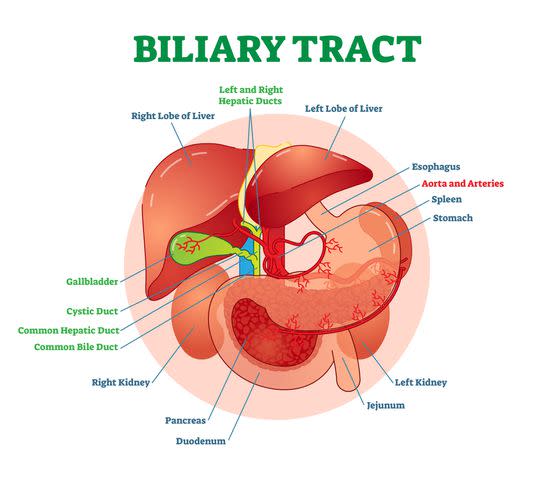
Biliary System Anatomy
The organs, ducts, and other structures of the biliary system are located in the upper-right abdominal quadrant, while the gallbladder is located just below the liver.
The extrahepatic ducts are located outside of the liver and are connected to the liver and gallbladder. Their function is to transport bile. Note that some bile ducts are also located inside the liver. These drain bile out of the organ and are called intrahepatic ducts.
Structure
The biliary system is comprised of a series of ducts, organs, and other structures responsible for producing, storing, and transporting bile. The bile is made in the cells of the liver and travels to the gallbladder to be stored for later use.
When you eat a fatty meal, the bile is released and travels to the small intestine through this system of ducts to its final destination, the duodenum.
Bile Flow Through the Biliary System
Bile travels in a controlled manner through the system of ducts and other structures of the biliary system.
Bile is made in the liver cells and flows from the liver into a system of ducts located inside and outside of the liver. These ducts function to collect the bile. Once collected, the bile travels to the right and left hepatic ducts.
It then flows from the right and left hepatic ducts into the common hepatic duct, then into the cystic duct, which is connected to the gallbladder.
From the cystic duct, the bile flows into the common bile duct (CBD). The common bile duct (CBD) is located where the common hepatic duct and the cystic duct join. It runs from the liver through the pancreas and into the duodenum.
The lower portion of the CBD joins the pancreatic duct before entering the duodenum. This is where pancreatic juices containing digestive enzymes enter the biliary system.
Bile is excreted into the duodenum through a muscular opening called the sphincter of Oddi.
The sphincter of Oddi relaxes to allow bile to enter the duodenum. Once the bile enters the duodenum, it begins to break down ingested fats. Only half of the bile ends up in the duodenum, while the other half travels into the gallbladder through the common bile duct.
The bile received by the gallbladder is stored in the gallbladder for future use.
Once bile is stored in the gallbladder, it isn’t released until a large meal is eaten and a hormone called cholecystokinin is secreted. This hormone stimulates the release of bile to begin the process of breaking down fats.
Anatomical Variations
Aberrant ducts are a common variation from the normal anatomy of the biliary system. Aberrant ducts are not anatomically structured the way they should be. For example, the ducts may abnormally join the wrong ducts, so that bile does not flow properly.
In fact, according to a 2006 study published in Liver and Biliary Tract Surgery, “50% of patients presenting with gallbladder stones or common bile duct stones show a significant variation from what is generally considered as the expected normal pattern.”
A 2011 study discovered as many as 22 variations of bile ducts in 59.5% of the study participants who had liver surgery. These included an extra right hepatic duct (in which a cystic duct drained) and five other abnormalities that had never been described before.
Variation from the normal anatomy of the bile ducts is a primary reason the ducts get inadvertently injured during some types of surgery.
Associated Conditions
Biliary disease describes any condition that affects the gallbladder, bile ducts, and other structures needed to produce and transport bile. Common maladies of the biliary system include gallbladder disease, biliary colic, and bile duct obstruction.
Gallbladder Disease
Gallstones are the most common gallbladder condition, but tumors and acute acalculous cholecystitis (sudden, severe inflammation of the gallbladder without gallstones) are other common types of biliary disease.
Related: Gallbladder Disease
Biliary Colic
Biliary colic is intermittent pain in the upper-right quadrant of the abdomen or above the stomach (epigastrium). Caused by a temporary obstruction of the cystic duct (this is usually secondary to a gallstone that is trapped in the cystic duct), the pain resulting from this condition can range from mild to severe.
If the obstruction is not removed or the gallstone doesn’t pass on its own, it results in cholecystitis (an acute inflammation of the gallbladder).
Related: An Overview of Biliary Colic
Bile Duct Obstruction
Also known as biliary obstruction, this is the blockage of any of the ducts in the biliary system. This condition most commonly occurs from a gallstone, but can also be caused by a tumor or another underlying cause.
Treatment
Treatment for biliary disease may include:
Medications to increase the flow of bile from the liver.
Antibiotics to treat an infection.
Hepatoportoenterostomy: A surgical procedure to drain bile from the liver when bile ducts are blocked.
Endoscopic retrograde cholangiopancreatography: A minimally invasive surgical procedure performed by a gastroenterologist. An endoscope (a flexible tube with a light and a camera) is used to locate and remove gallstones from the bile duct.
Cholecystectomy: Surgical removal of the gallbladder.
Tests
Several types of tests are done to diagnose abnormalities and diseases of the biliary system.
Liver function tests: A blood sample is taken, and a lab test is performed to evaluate certain enzymes and protein levels to see how the liver is functioning.
Endoscopic ultrasound: An endoscope is inserted through the mouth into the digestive tract. High-energy sound waves (ultrasound) are bounced off the endoscope, creating an image of body tissues.
Computed tomography (CT) scan: CT involves taking a series of images at different angles inside the body.
Magnetic resonance imaging (MRI): MRI uses radio waves and magnetic fields to create a series of detailed images inside of the body.
Endoscopic retrograde cholangiopancreatography: Used for treatment (see above), this procedure is also used to diagnose problems in the biliary system. It is sometimes combined with MRI, in a procedure called magnetic resonance cholangiopancreatography, to detect gallstones and diagnose the cause of other obstructions in the biliary tract.
Liver biopsy: This procedure involves the removal of a very small portion of liver tissue, which is examined in the lab for signs of disease or damage.
Summary
The biliary system includes the series of ducts and organs that produce, secrete, and store bile. Bile is a greenish-brown substance that helps break down fats and absorb nutrients. Bile is produced in the liver and stored in the gallbladder. It is released when you eat a fatty meal.
Gallstones can cause problems in the biliary system when they block bile ducts. Conditions of the biliary system are usually diagnosed with liver function tests and/or imaging tests or biopsy.

