20 Types of Skin Lesions and What They Look Like
Examples of benign and potentially cancerous lesions
Medically reviewed by Mary Choy, PharmDFact checked by Nick Blackmer
Skin lesions are abnormal changes in the skin compared to the surrounding tissue. Skin lesions may look like bumps or patches, or they may be smooth. They may be a different color or texture compared to nearby skin.
Common causes include acne, cellulitis, and chickenpox. This article looks at 20 types of skin lesions, their causes, pictures, and their treatment.
What Are Skin Lesions?
The term “skin lesion” refers to any skin area that is different from the surrounding skin.
Skin lesions may be raised like a cyst or flat like an age spot. They can be symmetrical like a wart or asymmetrical like melanoma. Some lesions, like skin tags, are the same color as your skin, while other lesions are darker, such as a mole.
There are many different types of skin lesions that you can be born with or acquire. Some are benign, which means they are harmless. Others can be severe and cancerous. They may appear all over your body, like hives, or they may be in one place.
The way a skin lesion looks and where it appears can help identify it. To find the cause of a skin lesion, healthcare providers consider:
Color
Size
Texture
Location
Types of Skin Lesions, With Pictures
Acne
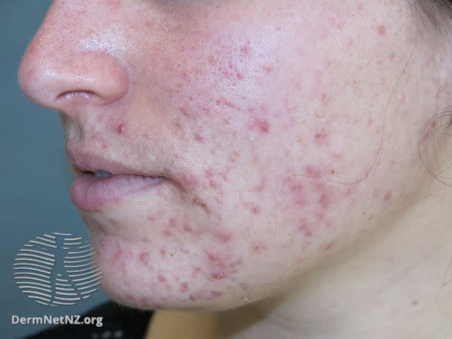
DermNet / CC BY-NC-ND
Acne presents primarily as papules. It can also cause pustules, nodules, or cysts. Acne is most common on the following places:
Face
Neck
Chest
Back
Acne can leave scars if not treated.
Acne occurs when your pores become blocked with dead skin or an oily secretion called sebum. It can also be caused by too much of a certain kind of bacteria on the skin.
Actinic Keratosis
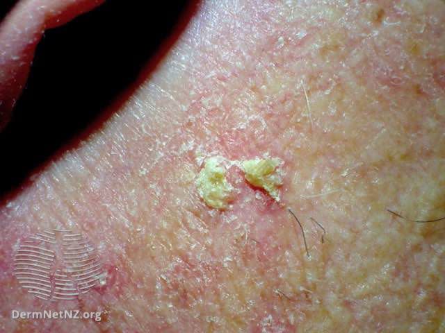
DermNet / CC BY-NC-ND
Actinic keratosis is caused by exposure to sunlight. It appears as thick, scaly crusts on the skin. It is most common in people over the age of 40. Because it is related to years of sun exposure, it doesn’t usually appear until later in life.
Actinic keratosis has a scaly, raised surface. This makes it easier to feel than see. Over time, it can turn hard and wart-like. It may develop a horn-like texture.
Get It Checked
Left untreated, up to 10% of actinic keratoses will develop into squamous cell skin cancer. About 40% to 60% of squamous cell cancer cases begin as actinic keratosis. If you think you might have actinic keratosis, see your healthcare provider. Identifying and treating it early can help prevent skin cancer from developing.
Blisters
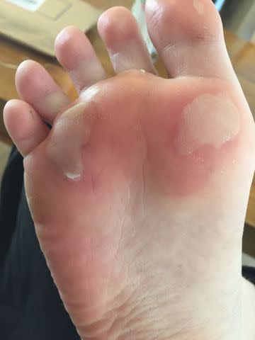
Johnny Eickelman / Getty Images
Blisters are fluid-filled areas of the skin. They can be caused by friction, such as a shoe rubbing on your skin when you walk. They can also be due to a burn, skin disorder, or allergic reaction.
Most blisters can be treated at home. Try not to pop blisters. Avoid putting more pressure on the blister area.
If a blister does pop, clean the area. Cover it with a sterile bandage until healed.
See a doctor if you have signs of infection whichinclude:
Pus, a thick yellowish or greenish discharge
Fever
Red hot skin around the blister
Takeaway
When a blister is less than 0.5 cm, it is called a vesicle. When it’s greater than 0.5 cm, it is called a bulla.
Cellulitis
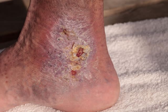
LagunaticPhoto / Getty Images
Cellulitis is a skin infection. It happens when bacteria or fungi enter the skin.
Symptoms of cellulitis include:
Redness
Swelling
Pain
Leaking of fluid
Cellulitis feels hot to the touch. It can also cause a fever and you may feel unwell.
If identified early, most cases can be treated at home with antibiotics. If left untreated, severe and serious complications can occur. Sometimes hospitalization may be needed.
When to Seek Medical Attention
It is important to see a doctor if you have signs of cellulitis. If you notice areas of swelling, redness, pain, or heat in your skin, especially where you’ve had a cut, bite, or burn, seek medical care at once.
Chickenpox
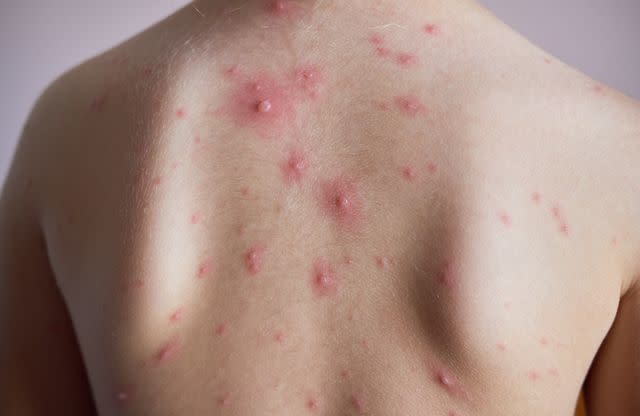
Alex Tihonovs / Getty Images
Chickenpox, also called varicella, is a viral skin infection.
Chickenpox causes red, fluid-filled blisters all over the body. They are typically hot and itchy. The blisters can ooze pus and may cause secondary crusting.
Chickenpox is contagious two days before the rash first appears. It remains contagious until all the blisters have crusted over.
It is possible to recognize chickenpox at home. However, you may want to see a healthcare provider to confirm the diagnosis.
Comedones
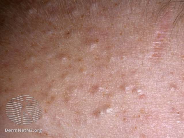
DermNet / CC BY-NC-ND
Comedones are a kind of acne.
There are a few different types:
Blackheads are open comedones, which means the pore is still open. They have a dark spot in the middle.
Soft closed comedones are soft, painless, and smooth.
Hard closed comedones have white heads. They are also called milia. They are not the same as whiteheads, which are filled with pus.
Cold Sores
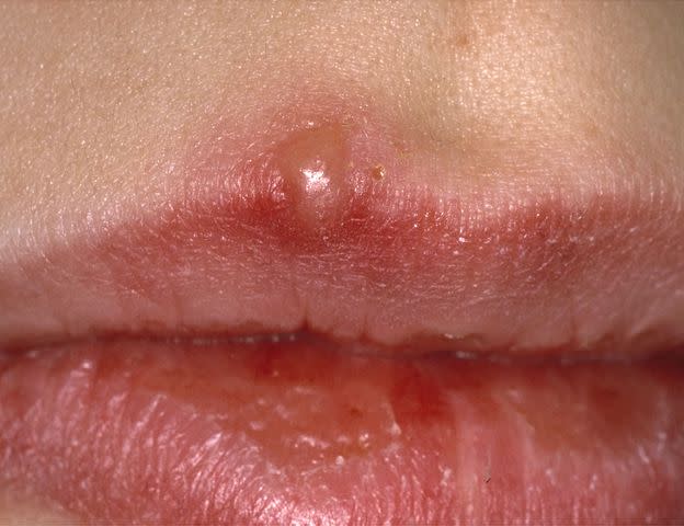
DermNet / CC BY-NC-ND
Cold sores are sores on the mouth or lip area. They are caused by the herpes simplex virus (HSV).
Cold sores can be red, painful, and cause fluid-filled blisters. You may feel tingling before the sore appears. Because they are caused by a virus, you may also have flu-like symptoms.
Very severe cold sores or sores that don’t seem to be healing on their own may require treatment from a healthcare provider.
HSV-1 vs. HSV-2
There are differences between HSV-1 and HSV-2:
HSV-1 usually causes oral herpes. Cold sores and fever blisters appear around the lip and mouth area.
HSV-2 is usually responsible for genital herpes.
However, oral or genital outbreaks may be caused by either virus.
Contact Dermatitis
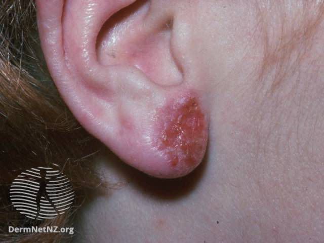
DermNet / CC BY-NC-ND
Contact dermatitis is caused by an allergen or substance that irritates the skin. Symptoms usually appear just in the area that contacts the irritant.
Symptoms may include:
Redness
Itching
Macules
Papules
Fissures
Blisters
Plaques
Swelling
Tenderness to the touch
If contact dermatitis does not get better at home, see a doctor.
Eczema
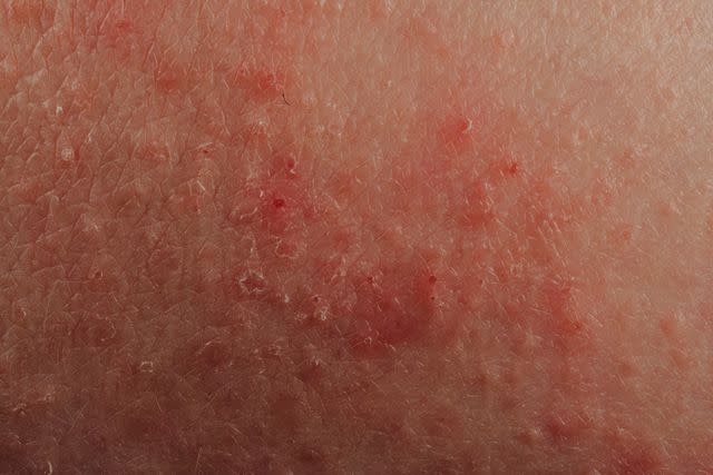
Pan Xunbin / Getty Images
Eczema is also called atopic dermatitis. It appears as an itchy, red rash. Symptoms may include:
Red, grey, brown, or yellow patches of skin
Itching
Dryness
Blisters
Fissures
Plaques
Sensitive and painful patches
Certain external conditions like hot or cold weather can cause symptoms to flare up. Some skin products may also contribute to irritation. Food allergies, hormonal changes, and pet/dust mites can often worsen symptoms.
The most common treatment types include:
Keeping skin hydrated with an emollient like a moisturizer
Topical soaps and creams
Corticosteroid cream
Avoiding triggers and irritants
Mild eczema can be managed at home with over-the-counter remedies. If you have more severe eczema, your primary care provider or dermatologist can help you with a treatment plan.
Related: Feet Eczema: Treatment, Triggers, and Lifestyle Changes
Freckles

Talia Ali / Getty Images
Freckles are small, flat, light-brown macules on the skin. They are caused by sun exposure.
Most common freckles are harmless and rarely turn into skin cancer. They are more common in people with light, fair skin, or red hair.
Freckles do not require any treatment unless they change and skin cancer is suspected.
Ephelides and Solar Lentigines
Two types of lesions commonly called freckles include:
Ephelides: These are typical freckles that occur from childhood. They are more common in people with fair skin and/or red hair. They tend to be round in shape and just millimeters in size. They appear in various brown shades.
Solar lentigines: These are macules with clear borders. They vary from light brown to black. They are most often called sun spots, liver spots, age spots, or actinic lentigines. They are caused by chronic sun exposure and are most commonly seen in older individuals.
Hives
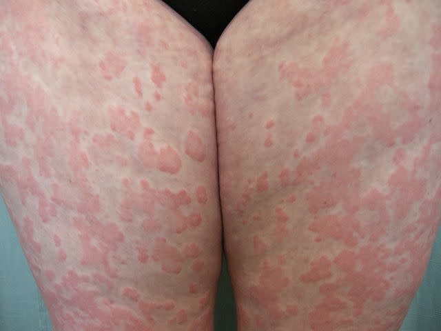
Raimo Suhonen / DermNet / CC BY-NC-ND
Hives are also called urticaria. They look like red, swollen, raised marks on the skin.
Hives are usually caused by an allergic reaction to something specific. They can also happen for unknown reasons.
Hives can itch or sting. Typically, the individual marks last less than 24 hours, though more can continue to appear.
Urticaria vs. Angioedema
Urticaria and angioedema are very similar. Urticaria only affects the skin, though, and each mark lasts less than 24 hours. Angioedema may last for days.
Urticaria occurs on the outer layer of the skin. Angioedema occurs under the skin. Angioedema can also affect the mucous membranes, such as the eyelids and lips. It can be painful. It often presents as deep swelling around the mouth and eye areas. It can sometimes affect the genitals, hands, or feet.
Severe angioedema can cause the throat or tongue to swell. This may create breathing difficulties. It may also cause swelling of the intestinal tract lining, which can lead to gastrointestinal cramping.
It is possible to get both urticaria and angioedema at the same time. Treatment for both conditions is similar. If breathing is affected, it is essential to seek emergency medical attention immediately.
Whether it is urticaria or angioedema, call seek emergency medical care if you experience:
Difficulty breathing
Wheezing
Chest tightness
Tongue/facial swelling
Dizziness
Impetigo
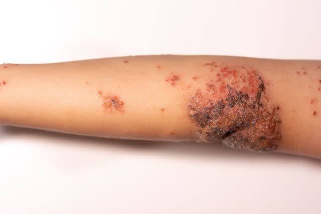
Matthew Roberge / Getty Images
Impetigo is a bacterial skin infection. It is caused by bacteria entering the skin through a hair follicle or a small cut. The condition causes sores, crusts, and blister-like bumps.
Impetigo is very contagious. It most commonly affects young children.
Keloids

wutwhanfoto / Getty Images
Keloids are raised scars that form after an injury. They range in color from flesh-colored to red. Keloids are caused by an overgrowth of scar tissue and are often itchy and uncomfortable.
Some people are more prone to developing keloids. However, certain preventative measures may stop keloids from forming.
Those who have had keloids in the past have a higher risk of developing them again. To prevent this, there are a few things you can do:
Pay close attention to any thickening skin in injured areas. If you notice this after an ear piercing, for example, immediately remove the earring and wear a pressure earring over the piercing.
Practice good wound care. Wash with soap and water and bandage with sterile petrolatum gauze. Apply silicone gel or silicone sheets to freshly healed scars.
Keloids are harmless and do not require medical attention. Still, some people may feel distressed by the way they look. If you notice any thickening of the skin around a recently healed wound, consult your dermatologist. Treatment can help reduce their appearance.
Related: Piercing Bump vs. Keloid: What Are the Differences?
Moles
Skin Cancer Foundation
A mole is also known as a melanocytic nevus or simply a nevus. They are usually round, brown/pink macules, papules, or nodules. They can be found anywhere on the body and may appear at any age.
Moles are usually harmless. However, see a doctor if a mole changes shape, color, size, or begins to bleed or crust. This could be a sign of melanoma, a type of skin cancer.
ABCDE Rule
When checking your moles, these changes may suggest melanoma:
Asymmetrical: Look at the shape of the mole. Moles are usually even in shape and look similar on all sides. Melanomas tend to be uneven in shape.
Border: Normal moles have a smooth edge. Melanomas typically have an irregular, jagged border.
Color: Moles tend to be even in color throughout. Melanomas might be uneven in color.
Diameter: Moles tend to be small. Melanomas are usually over 6 millimeters wide.
Evolving: Moles don’t tend to change much. Melanomas often change in size, shape, color, or texture.
Keep an eye out for changes in your moles or new moles with these qualities. If you find anything suspicious, see a healthcare provider right away.
Related: How to Tell If a Mole Is Turning Into Skin Cancer
Psoriasis
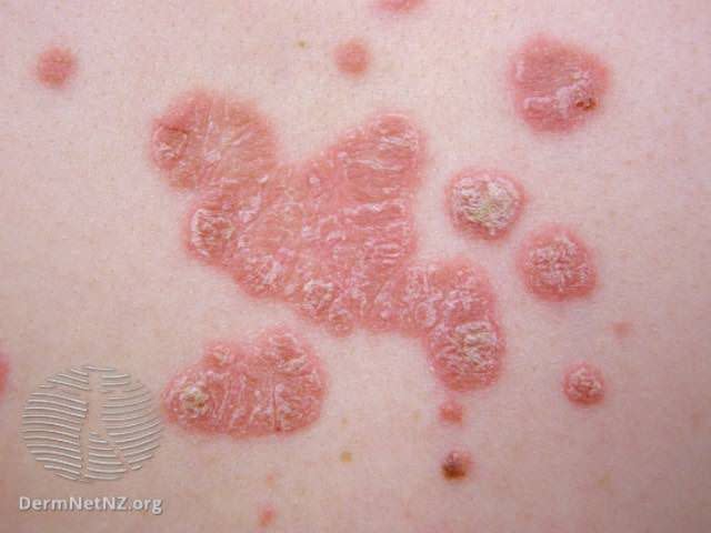
DermNet / CC BY-NC-ND
Psoriasis tends to look like red, flaky skin, crusty patches of skin, and silvery skin scales in fair-skinned people. Hispanic people are more likely to have salmon-colored psoriasis and silvery-white scales. In African Americans, psoriasis often looks violet and the scales are gray.
Psoriasis happens because the body produces skin cells too quickly, over days rather than weeks. The excess growth of skin cells causes thick, raised, scaly patches.
The most common areas for psoriasis to occur are:
Knees
Elbows
Scalp
Lower back
Still, it can occur anywhere on the body. There is no cure for psoriasis.
Eczema vs. Psoriasis
Eczema and psoriasis look similar. There are a few differences to look out for:
Itching tends to be much more intense with eczema and milder in psoriasis.
Eczema tends to occur in places like the crooks of the knees and the elbows. Psoriasis most commonly occurs on the elbows, knees, scalp, and lower back.
With eczema, the skin tends to be red, inflamed, cracked, blistered, and may leak pus. With psoriasis, the skin appears scaly, flaky, thickened, raised, and can be silvery.
If you are unsure if you have eczema or psoriasis, consult a doctor or dermatologist. The correct diagnosis will help ensure you get the right treatment.
Related: What May Be Causing Pimples on Your Elbow
Scabies
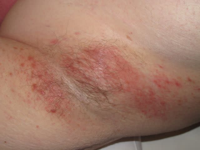
DermNet / CC BY-NC-ND
Scabies is caused by a mite. It begins with intense itching. A rash can also develop. The rash appears as small red papules, welts, or scaly lesions. Repeated scratching can damage the skin.
Sebaceous Cyst

Sebaceous cysts are round and filled with keratin, a protein found in skin, hair, and nails. The nodules are firm and usually skin-colored. They usually appear on the upper body or face. They can range in size and occasionally rupture.
A ruptured cyst may become infected and needs to be treated. Otherwise, sebaceous cysts don’t usually require treatment. They tend to grow slowly and are benign.
Shingles

Shingles is caused by the reactivation of the virus that causes chickenpox. A shingles rash is a very painful red rash comprised of macules, papules, and blisters. The blisters can break and weep fluid. This can lead to secondary crusting.
The rash itches, burns, and can be painful. Other symptoms can include:
Fever
Chills
Headache
Lethargy
You may hear shingles referred to as a belt or band. This is because it often appears as a belt-like formation around the rib cage or waist.
Takeaway
The risk of shingles can be reduced with vaccines like Shingrix. The Centers for Disease Control and Prevention (CDC) recommends the Shingrix vaccine for people over 50 and for younger adults who have weakened immune systems. The vaccine also protects against postherpetic neuralgia, a painful condition of the nerves. This is the most common complication of shingles.
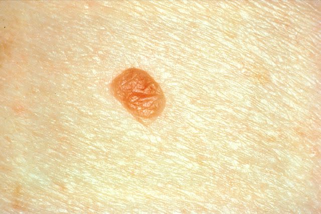
Skin Tag
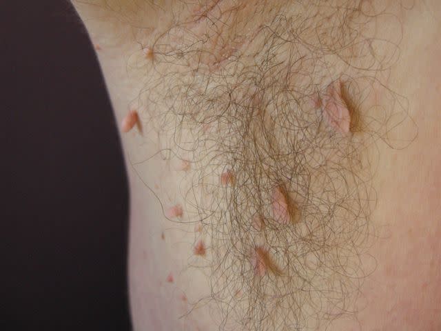
DermNet / CC BY-NC-ND
Skin tags are also called acrochordon. They are soft, small, skin-colored growths and occur more often as people age.
Removal is not usually necessary. Skin tags usually don’t require medical attention unless:
They cause cosmetic concern.
They’re in a position that causes irritation.
Related: Can You Cut a Skin Tag Off at Home on Your Own?
Warts
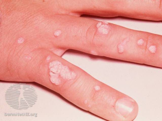
DermNet / CC BY-NC-ND
Warts are caused by the human papillomavirus (HPV). They tend to occur in groups and are contagious.
Warts are raised, flesh-colored papules. They may contain tiny black dots in the center. They are not dangerous but can be painful and are easy to pass to others.
There are several different types of warts, including:
Common warts
Filiform warts
Genital warts
Periungual warts
Takeaway
Most warts are not dangerous. Rarely, some types of human papillomavirus that cause genital warts can cause cervical or anal cancer.
Primary vs. Secondary Skin Lesions
Skin lesions are either primary or secondary. Primary skin lesions are either present from birth or develop during your lifetime.
Secondary skin lesions arise from primary skin lesions. This can happen when a primary skin lesion is:
Disturbed
Irritated
Changes over time
For example, if eczema is scratched, a crust may form. The crust is a secondary lesion.
Primary Skin Lesions
Primary skin lesions tend to be divided into three groups:
Lesions formed by fluid within the skin layers: Examples include vesicles and pustules.
Lesions that are solid masses: Examples include nodules and tumors.
Flat lesions: Examples include patches and macules.
Types of primary lesions include:
Bulla: A vesicle that is more than 0.5 centimeters (0.2 inches) and filled with fluid
Cyst: A raised area of the skin that has clear borders and is filled with fluid or semi-solid fluid
Macule: A flat, discolored lesion, less than 0.5 centimeters (0.2 inches) in size
Papule or maculopapular: An elevated solid lesion, up to 0.5 centimeters (0.2 inches) in size. It has clear borders, is firm, and can appear in various colors.
Patch: A flat, discolored lesion, greater than 0.5 centimeters (0.2 inches) in size
Plaque: A lesion that is raised like a papule, greater than 1–2 centimeters (0.4 to 0.8 inch) in size. It is solid, rough, and flat-topped.
Vesicle: A fluid-filled blister less than 0.5 centimeters (0.2 inches) in size
Pustule: Similar to a vesicle but filled with pus instead of fluid
Nodule: A circular, elevated, solid bump greater than 0.5 centimeters (0.2 inches) in size
Telangiectasia: Clusters of “spider veins” where tiny blood vessels cause red lines on the skin
Tumor: A lesion larger than 0.5 centimeters (0.2 inches) in size that looks similar to a nodule. Tumors can be benign or cancerous.
Wheal: An irregular-shaped, solid, elevated area that can vary in color and lasts for only a short time
Secondary Skin Lesions
Examples of secondary skin lesions include:
Atrophy: Skin that is paper-thin, transparent, and wrinkled. It is usually due to the use of a topical medicine like steroids.
Crust: A rough, elevated area formed from dried fluid. The fluid may be pus, blood, or serum.
Erosion: The loss of the top layer of skin. It is moist and glistening in appearance.
Excoriation: Straight scratches that result in the loss of the top layer of skin
Fissure: Straight breaks in the skin that go deeper than the top layer of skin into the second layer of skin. They can be painful and can be caused by excessive dryness.
Lichenification: A rough thickening of the top layer of skin
Maceration: Skin that is wet, wrinkly, and lighter in color. This happens when skin is in contact with water or fluid for too long. It can occur due to leaking wounds that have been improperly cared for.
Phyma: A thickening of the skin. This is often seen in advanced rosacea.
Scale: A build-up of cells that form patches and then flake off the skin
Ulcer: A wound deeper than the top layer of skin that damages the second layer of skin. It is concave and variable in size. Deeper ulcers are more serious.
Umbilication: A dip inside a skin lesion that looks similar to a navel.
How Are Skin Lesions Diagnosed?
If you notice a lesion on your skin, schedule an appointment with your healthcare provider or dermatologist to have it looked at. During your appointment, they will physically examine the characteristics of the lesion, including its size, shape, color, texture, and location.
Your provider will ask you questions about the lesion, like when you noticed the change in your skin, and whether or not the lesion itches. You can also expect them to inquire about any allergies or health conditions that you have, medications you are taking, and other pieces of your medical history.
Healthcare providers are often able to diagnose skin lesions simply by looking at them. But they may order additional tests to confirm a diagnosis, including:
An allergy test, such as a skin prick test, to check for hypersensitivity to specific allergens
A blood test, such as an immunoglobulin E (IgE) blood test, to check for immune system activity that could point to a skin disease or infection
A microbial swab, to determine which, if any, bacteria or fungus is causing the lesion
A skin biopsy, to diagnose or rule-out malignancy (cancer)
How Are Skin Lesions Treated?
Most skin lesions are benign (harmless) and don’t need to be removed unless for cosmetic reasons. Your healthcare provider may recommend having the lesion removed if it is causing you discomfort or if there is any doubt that it could be cancerous.
Skin lesion removal can typically be done in your healthcare provider’s office, although in some cases it may need to be removed by a surgeon. The lesion and surrounding skin will likely be numbed with a local anesthetic before the removal.
Your healthcare provider may use one of the following methods to remove the lesion:
Shave excision is a technique used for raised lesions or lesions in the upper layer of skin. A provider will remove all or part of the lesion using a small blade. Stitches are usually not necessary.
Simple scissor excision is another method used for raised lesions or those in the upper skin layer. The provider will use forceps to gently pull the lesion up from the skin, then cut around the lesion with scissors. Stitches are usually not necessary.
Skin excision (full thickness) is a method used when there is a concern about skin cancer. The entire lesion is removed down to the fatty layers of skin, and some of the surrounding tissues may be removed to ensure the entire area is clear of cancer cells. The area is closed with stitches.
Laser excision is used to remove benign lesions, such as warts, moles, and even tattoos. The provider will aim a specialized laser beam at the lesion, heating up the skin pigments until they burst and can be carried away by the immune system. Stitches are not needed.
Cryotherapy is a method used for warts, actinic keratoses, and seborrheic keratosis, in which liquid nitrogen is applied to the lesion to freeze it. Following the procedure, the lesion will blister and peel away.
If diagnostic tests reveal that the skin lesion is malignant, surgery to remove as much of the affected skin as possible in order to prevent it from growing back. Depending on the severity of the malignancy, radiation therapy and/or chemotherapy may be necessary to destroy any remaining cancer cells.
Summary
Skin lesions can be present from birth or develop over your lifetime due to sun exposure, bacteria, allergies, or chronic conditions. They can vary in appearance and may or may not be cancerous.
Many lesions can be treated at home with topical ointments and creams. Others require treatment from a healthcare provider. If you are unsure of what type of lesion you have, or if you have a lesion that changes suddenly, it is always best to seek medical care.
Related: Common Skin Diseases and Conditions
Frequently Asked Questions
What kind of doctor treats skin lesions?
Ongoing skin conditions like acne, eczema, or psoriasis often require treatment by a dermatologist, or skin specialist. Others, like impetigo, can be treated by your primary care provider. And some—like blisters and chickenpox—go away on their own.
Are skin lesions usually cancerous?
Not usually. However, some types of lesions can become cancerous. One example is actinic keratosis, a rough, scaly patch or bump on the skin caused by UV damage. Actinic keratosis lesions can turn into squamous cell skin cancer. This type of lesion is often called “precancerous.”
What does a cancerous lesion look like?
Signs of cancerous skin lesions include any changes in the skin, including new growths, changes to existing growths, and growths that have difficulty healing.
Read the original article on Verywell Health.

