Students create realistic 3D models of human organs in new class at UNF
Wearing blue rubber gloves, Molly Dobrow reached into a metal vat of diluted sodium hydroxide and pulled out two dripping-wet models of human organs: a heart and a set of lungs.
Made of Elastico, a kind of elastomer, they had been 3D printed in a lab at the University of North Florida, then put in the sodium hydroxide bath, which ate away any unneeded material, leaving these scaled-down replicas that recreate human organs and body parts with remarkable accuracy.
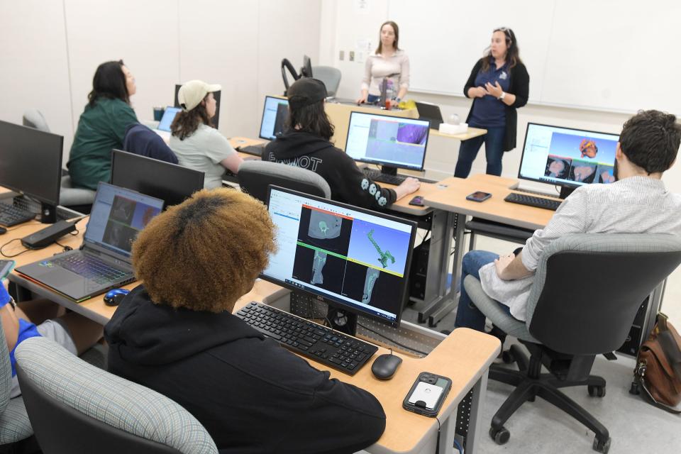
It's all part of a UNF undergraduate class called Anatomy in 3D taught by Laura Habegger, assistant professor of biology, and Grant Bevill, associate professor of mechanical engineering.
The university teamed up with Mayo Clinic to offer the class for the first time this spring semester.
The course gives biology and engineering students experience in a growing medical field. Habegger said it could help students find jobs, if they choose such a path, in 3D medical printing labs.
More medical professionals are using 3D printing to create duplicates of a patient's organ based on numerous CT and MRI scans. Surgeons can then, before even making the first incision, examine the organ and map out their surgical approach, Habbenger said. That can make for better outcomes and reduced time in surgery. Some insurance companies are even starting to cover the cost of models, she said.
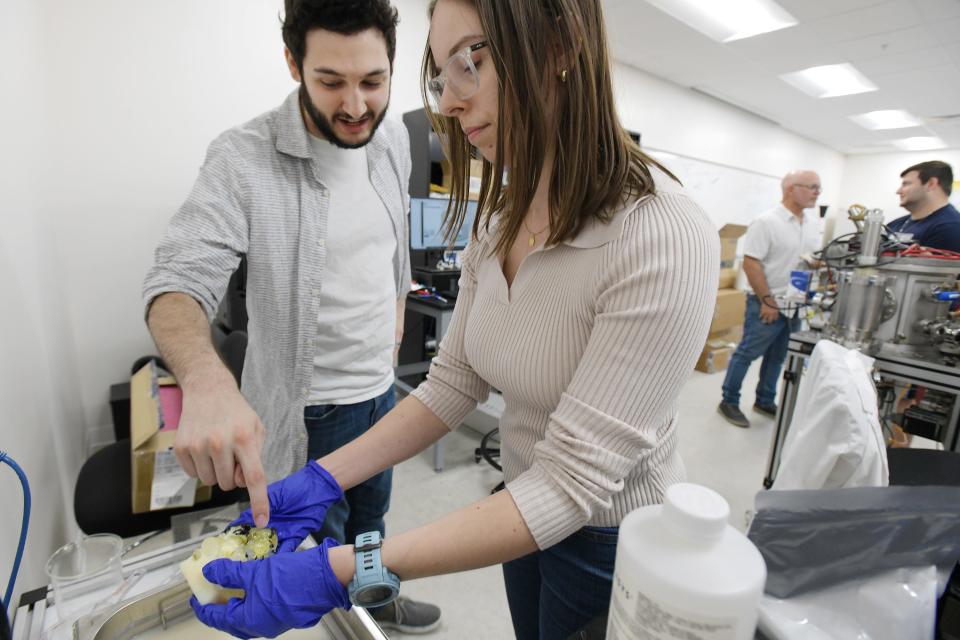
Dobrow, a graduate assistant, displayed a model of a brain made in yellow Elastico, inside of which was a small mass of black material that represented a brain tumor — all based on the scans of an anonymous patient.
"It's very common in surgical practice for surgeons to request the development of these models so they can do surgical preplanning," said Dobrow, who has been accepted into the University of Florida's doctorate program in biomedical engineering. "The surgeon can actually practice."
Using some 600 slices of CT scans each, students Ansley Bates and John McGarity had created spines showing a patient with scoliosis — both before surgery (Bates' model, with a sharp bend) and after (McGarity's, straighter with implanted rods to support it).
No 'ick' factor: JU's 3D learning tables provide cadaver-free method to learning anatomy
The students wore big grins as they saw the finished products. "You really get a better idea of anatomy from looking at this," Bates said.
Yooyeon Jung created a model of a brain with a tumor that you take out, hold and look at from all sides. "You could hold it in your hands," she said. "It really helps to observe it from all angles."
Jung got a degree in biology and is now getting a master's in mechanical engineering. The UNF class was designed to attract students in both of those fields to encourage collaboration and shared expertise.
Consider the project of Christian Diakos, a mechanical engineering student who wants to work in the field of prosthetics and medical devices, and Patrick Contiu, who's applying to medical schools this summer.
They were getting ready to print a model of the heart of an anonymous patient who has an implanted heart pump, as well as tuberculosis that's left extensive calcification on the heart — both factors that make it difficult for doctors to read conventional CT scans. Their heart model includes both the heart pump and the calcification.
Health care: New Baptist nurses get 'real-life' training at JU residency
Contiu said that teamwork with Diakos was essential. "He's the engineer, so he knew how to do all the computer work, and I came from the biological and anatomical aspect," he said.
"The beauty of an interdisciplinary class," Diakos said. "It is so cool."
This article originally appeared on Florida Times-Union: UNF class in Jacksonville creates 3D models of human organs

