Super-detailed map of brain cells that keep us awake could improve our understanding of consciousness
Scientists have charted a comprehensive map of the brain cells responsible for keeping us awake. In doing so, they aim to better understand the mechanisms that enable human consciousness and to improve treatments for people in comas and vegetative states.
"It's a beautiful study," said Dr. Nicholas Schiff, a professor of neurology and neuroscience at Weill Cornell Medicine who was not involved in the new research. "It's a map of everything."
Schiff, who has collaborated with some of the study's authors in the past, hopes the new map can serve as a template for scientists to go back and look at patients with altered states of consciousness. Such work could help clarify exactly how this wakefulness network can be disrupted and what's required to get it back online. It's a tool that could help scientists pinpoint which brain activity is "necessary and sufficient" to keep someone awake and aware, Schiff told Live Science.
The new study was published Wednesday (May 1) in the journal Science Translational Medicine.
Related: What happens in our brains when we 'hear' our own thoughts?
Mapping consciousness
Human consciousness arises from a delicate dance between the wrinkled surface of the brain, known as the cerebral cortex, and deeper "subcortical" structures beneath it. Circuits in the cortex direct our attention and process information from the world around us, while the subcortical networks plug into these circuits from below, essentially keeping them active.
"It sends the signals that activate and stimulate the entire rest of the brain," said first author Dr. Brian Edlow, co-director of Mass General Neuroscience and director of the Laboratory for NeuroImaging of Coma and Consciousness.
Thus, consciousness is a "seamless interaction of these subcortical networks with these cortical networks," Schiff said. "It's an artifice, I think, to try to separate these things."
But scientists' understanding of humans' subcortical networks is incomplete. It relies heavily on animal experiments, in which scientists can inject trackable chemicals into the brain and then remove the organ to get a good look at where they ended up. In living humans, these deep-seated networks are located near major arteries and air-filled sinuses, and they also move a bit with each breath, making standard brain scans less crisp and hard to decipher. By contrast, networks in the human cortex are easier to image and have been much better studied.
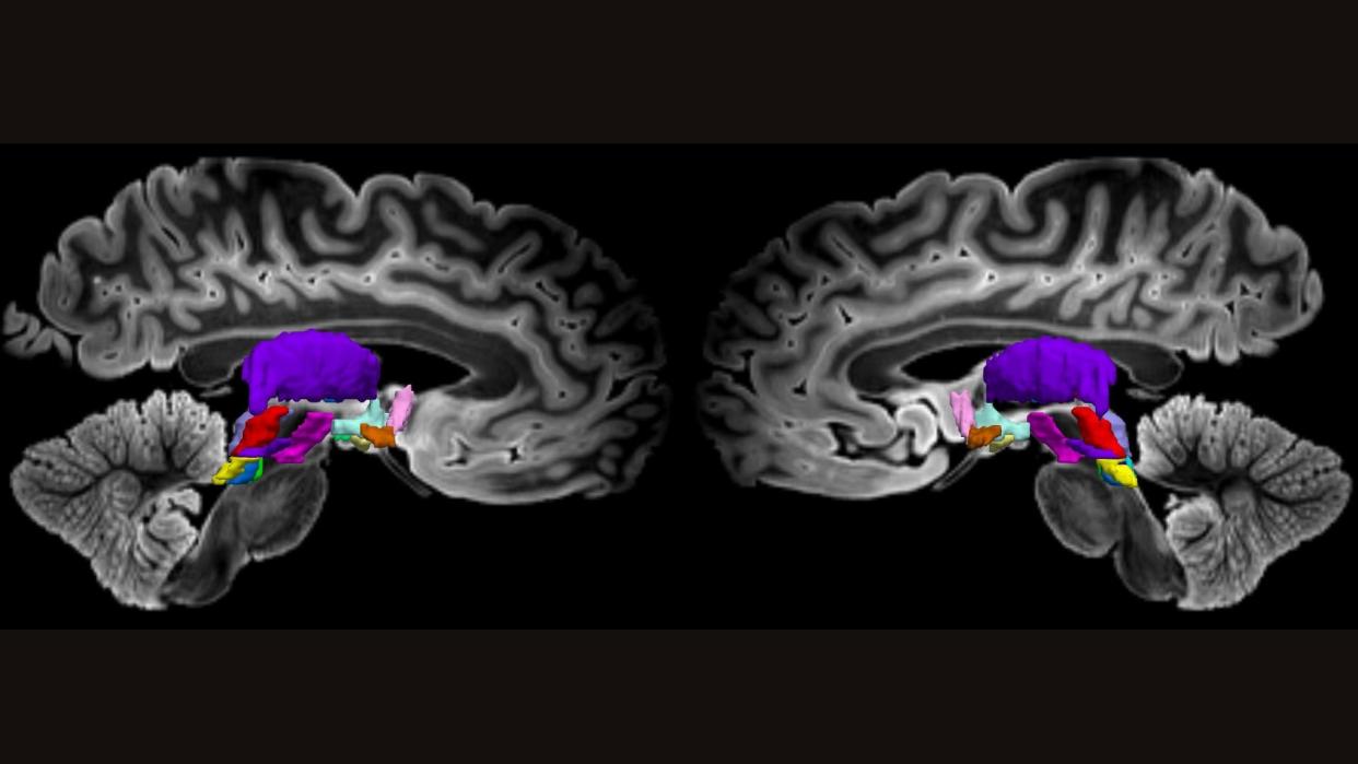
To help close this knowledge gap, the scientists behind the new study looked at brains from three middle-age organ donors. Using postmortem brains allowed the team to "map brain networks at ultrahigh resolution and to see connections that simply cannot be seen when you're scanning a living human being," Edlow told Live Science.
These organ donors had no neurological problems when they died, and their brains were carefully preserved in formaldehyde. The researchers stained the brains with colorful chemicals to mark different types of brain cells and then put the brains in an magnetic resonance imaging (MRI) scanner for up to 48 hours.
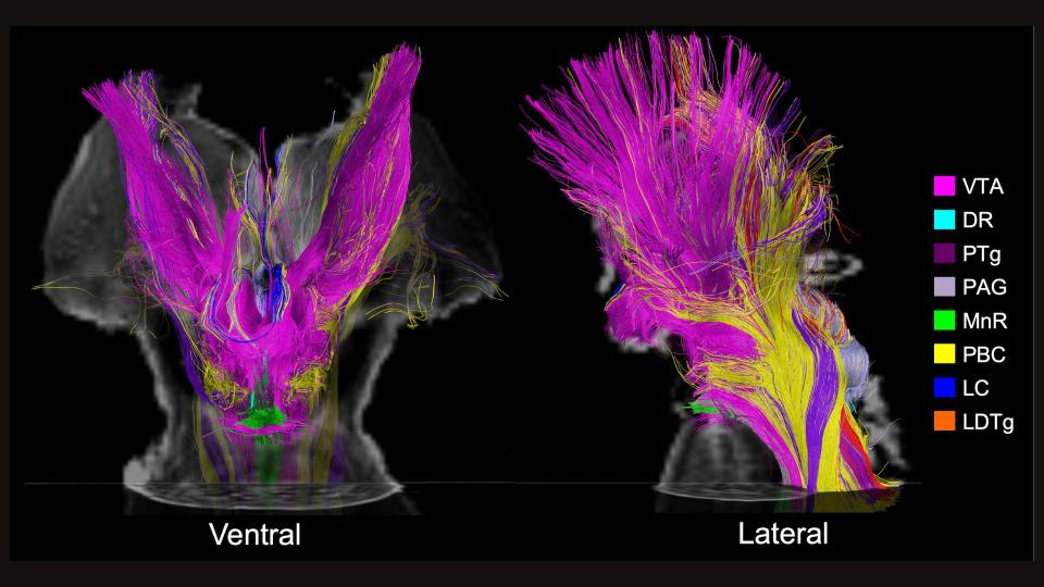
This resulted in incredibly high-resolution maps of subcortical structures that had been tied to wakefulness in previous research. The team manually traced connections between the brain cells in these structures, taking into account the direction water would flow down their wires. This metric captured by MRI can help reveal how strongly different nodes in a network are linked.
This work revealed major nodes of the wakefulness network in several subcortical structures — the brainstem, hypothalamus and thalamus — as well as part of the cortex called the basal forebrain. This network was confirmed to be linked to a circuit involved in awareness. Known as the default mode network, it is very active when we're daydreaming and not focused on a particular task.
Related: Ultrasound treatment 'jump-started' the brains of 2 people in coma-like state
To back up their work in postmortem brains, the team also looked at functional MRI data from more than 80 people who'd participated in the Human Connectome Project, a government-led effort to map all the networks in the human brain. These additional brain scans, which looked at the flow of oxygenated blood, pointed out a hub where these awareness and wakefulness networks meet: the ventral tegmental area.
RELATED STORIES
—Surges of activity in the dying human brain could hint at fleeting conscious experiences
—'Flow state' uncovered: We finally know what happens in the brain when you're 'in the zone'
—A mysterious brain network may underlie many psychiatric disorders
Taken together, the postmortem brain scans provided maps of highways in the brain, while the live scans showed traffic patterns along those roads. "We view our findings as an initial map that is beyond the spatial resolution that has been achieved previously," Edlow said. Looking to the future, the researchers hope to improve upon the map's resolution even more.
At this point, it's unclear exactly how the new map could shape the treatment of patients in comas, vegetative or minimally conscious states, Schiff noted. But it should be a useful tool to better understand and treat these conditions, he added.
"We envision that these connectivity maps will allow us to piece together, one individual at a time, the combination of connections that are necessary and sufficient to recover consciousness," Edlow said.
Ever wonder why some people build muscle more easily than others or why freckles come out in the sun? Send us your questions about how the human body works to community@livescience.com with the subject line "Health Desk Q," and you may see your question answered on the website!

