The first 3D, full-color X-rays will weird you out

Classic black and white x-rays, move down the bench.
New Zealand company MARS Bioimaging has developed the world's first full-color, 3D X-rays, and they're so real it's rather disturbing.
Developed over a decade by father and son scientists Phil and Anthony Butler in collaboration with the Universities of Canterbury and Otago, the MARS system is a new medical scanner using technology developed at CERN. And it could be more accurate than the typical scans you get a doctor's office these days.
SEE ALSO: This 'smart' prosthetic ankle makes it easier to use stairs
The MARS scanner uses a family of chips called Medipix, originally developed to track particles at the Large Hadron Collider. Medipix works like your camera — when the electronic shutter is open, each individual particle is detected and counted, creating high-res, accurate, noise-free images.
When used with the Butlers' MARS scanner and its software, the chips help to produce highly accurate, striking, three-dimensional color renderings of the human body that distinguish materials like metal, bone, soft tissue, and fat with different tones.
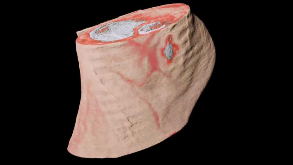
Here's a completely fascinating, if not somewhat horrifying video of an "ankle slice through," a series of 3D images taken by the MARS scanner. You can seen the skin and cartilage in beige, bone in white, and soft tissue and muscle in red.
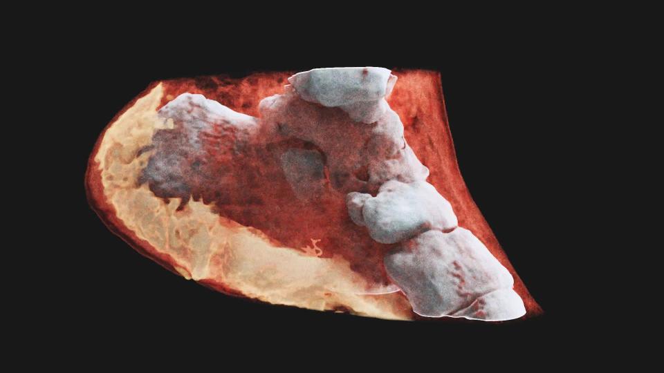
This one's also an ankle, this time in video rotation.
You can see the bones of the ankle, as the skin and muscles have been made translucent. You can also see the padding of the heel region, which looks super juicy and spongy — you're walking on this every day, guys.
It's all down to that Medipix3 chip, one of the most advanced chips available in the world, according to its makers.
"This technology sets the machine apart diagnostically because its small pixels and accurate energy resolution mean that this new imaging tool is able to get images that no other imaging tool can achieve," said Phil Butler in a statement published by CERN.
According to the company, MARS Bioimaging Ltd plans to commercialize the scanner in the future. So far, it's been used to study cancer, vascular diseases that lead to strokes and heart attacks, and bone and joint health.
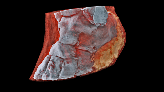
Image: MARS Bioimaging Ltd
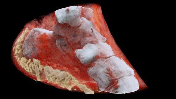
Image: MARS Bioimaging Ltd
“In all of these studies, promising early results suggest that when spectral imaging is routinely used in clinics it will enable more accurate diagnosis and personalization of treatment,” Anthony Butler said in the statement.
A group of orthopedic and rheumatology patients in New Zealand will be scanned in clinical trials over the next few months.
Just imagine getting your body parts scanned with this technology. If it's too real to think about seeing inside your own skin, just think how much more accurate this could be in comparison to regular black and white x-rays.
WATCH: This veteran firefighter and amputee is building powerful prosthetics for extreme sports