Chicago Bears linebacker Danny Trevathan has been suspended for two weeks after his illegal hit landed Green Bay Packers receiver Davante Adams in the hospital on Thursday.
Jon Runyan, the National Football League’s vice president of football operations, issued the decision on Saturday. Adams caught a pass midfield during the third quarter when he was hit by the Bears linebacker. Trevathan’s move had fans and commentators in an uproar, calling the dangerous helmet-to-helmet hit a “dirty” play.
A statement from the NFL’s communications office clarified that the unpaid suspension was due to Trevathan’s violation of Rule 12, Section 2, Article 6 (i) of the league’s player safety rules.
“There shall be no unnecessary roughness,” the statement reads. “This shall include, but not be limited to: (i) using any part of a player’s helmet or facemask to butt, spear, or ram an opponent violently or unnecessarily.”
Medical personnel took Adams off the field on a stretcher and he received preliminary exams for head and neck injuries at the stadium. He was later taken to the hospital for further testing. NFL Network’s Ian Rapoport reported that Adams suffered a concussion from the hit.
The Packers receiver tweeted on Friday that he was at home “feeling great.”
At home feeling great. Appreciate the prayers🙏🏾🙏🏾🙏🏾
As more studies reveal the prevalence of CTE, or chronic traumatic encephalopathy, in former football players, hits such as the one Adam are fueling calls for more rule changes in the National Football League. The degenerative brain disease has increasingly been found in postmortem tests on former players who had suffered repeated blows to the head.
Just like the electrical wires in the national grid, the electrical connections between brain cells, as shown in this picture, have to be well insulated. If this insulation is lost, neurons lose their ability to communicate efficiently. This is what happens in several neurological diseases including multiple sclerosis (MS).
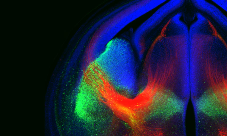
Navigating Axons: A Circuitous Route
This colorful picture shows the wiring in a developing brain. Axons (red) are the cables that neurons use to transmit their information, often over relatively long distances and taking highly circuitous routes. The other colors represent different areas of the brain.
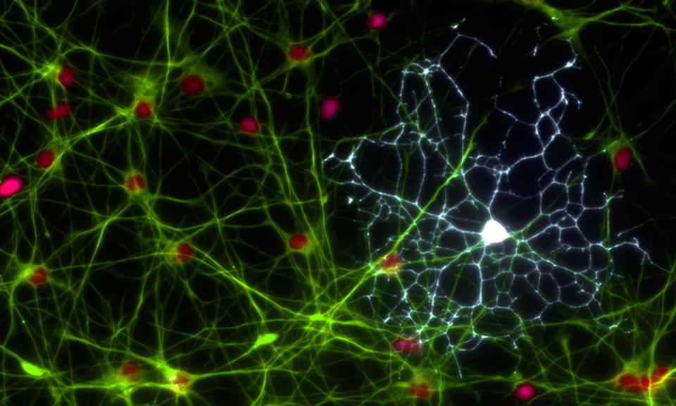
Glial Grandeur
At first glance this may look like a spider’s web but this web measures just 1/20 of a millimeter. It is made up of two types of brain cells – astrocytes in green and a white oligodendrocyte. These cells were originally thought of as the support cells for neurons but it is now known they are essential for many brain functions.
Female Mosaic
This picture of neurons from a female brain highlights those that have switched off the X chromosome inherited from the mother (in green), and those that have silenced the X chromosome inherited from the father (in red). In cases where an altered gene on one of the X chromosomes causes autism or intellectual disability, only around a half of the cells will be affected. This helps to explain why these conditions are less common in women than in men.
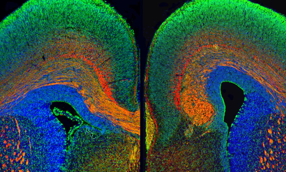
Breakdown In Communication
This image shows differences between a typical brain (left) and autism (right). The different colors identify different areas of the brain.
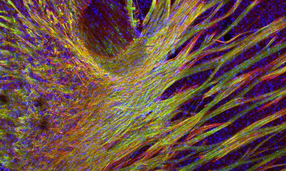
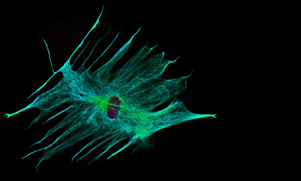
The Astrocyte
These star-shaped cells, or “astrocytes,” were once thought to be simple support cells for neurons. Now we know that they are much more important than this--they also help to create and maintain an environment in the brain that is optimized for electrical and chemical communication.
Neuron Networks
Scientists can use mathematics to model brain circuitry, as shown in this picture. They use this approach to predict how brain communication is altered in neuropsychiatric disorders, such as anxiety and ADHD.
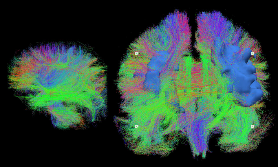
Wiring The Brain
This is a detailed map of the brain wiring in a sleeping newborn baby (left) and an adult in their seventies (right), visualized using MRI.
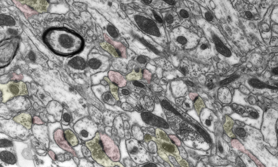
An Electron’s View Of The Brain
Neurons talk to one another across a gap called the synaptic cleft, rather than being directly connected to one another. A trained eye can identify the wires that are transmitting messages and those that are receiving information in this picture.
Circuit Building Block
Neurons have branched projections that extend from their cell body called dendrites which give the cells a tree-like appearance. It’s through these dendrites that neurons receive information from hundreds to thousands of other cells.
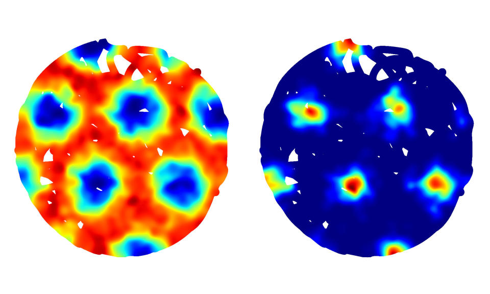
Encoding Space
Our brains hold specialized neurons called grid cells that help us to keep track of where we are. This heat map shows the regions in space where an individual grid cell becomes active during exploration of a circular room.
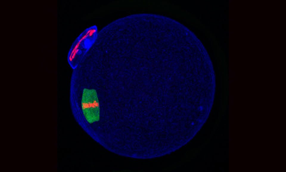
The Egg: Mendel's Moment
This picture shows the egg or “oocyte” preparing the genes that will be passed on to its offspring, which are highlighted in red.
Regenerating Spinal Cord
Images such as this one, which shows the spinal cord from a zebrafish repairing itself, are helping scientists to study biological mechanisms that could one day reveal treatments for people who are paralyzed due to spinal cord damage.
High Fidelity
This picture shows the difference in brain signals from a typical brain (left) and from a brain affected by a condition similar to Fragile X Syndrome, the most common inherited form of autism (right).
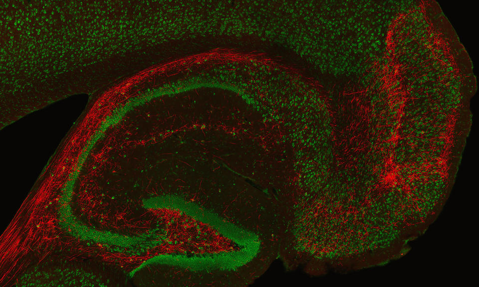
The Seahorse
This is a close-up image of a particular area of the brain called the hippocampus, named from the Greek word for “seahorse” because of its shape.
A Fragile Balance
This image shows a series of MRI pictures from the brain of an individual with Fragile X Syndrome, the most common inherited form of autism.
Former NBA guard Darius Morris has died at the age of 33. He played for five teams during his four NBA seasons. Morris played college basketball at Michigan.
Affluent Americans may want to double-check how much of their bank deposits are protected by government-backed insurance. The rules governing trust accounts just changed.
Miami Heat president Pat Riley rebuked comments Jimmy Butler made about the Boston Celtics and New York Knicks, while also implying that his star needs to play more.
Jake Mintz & Jordan Shusterman discuss the Padres-Marlins trade that sent Luis Arraez to San Diego, as well as recap all the action from this weekend in baseball and send birthday wishes to hall-of-famer Willie Mays.
An annual government report offered a glimmer of good news for Social Security and a jolt of good news for Medicare even as both programs continue to be on pace to run dry next decade.
Jason Fitz and Frank Schwab join forces to recap the draft in the best way they know how: letter grades! Fitz and Frank discuss all 32 teams division by division as they give a snapshot of how fans should be feeling heading into the 2024 season. The duo have key debates on the Dallas Cowboys, New York Giants, New Orleans Saints, Los Angeles Rams, New England Patriots, Las Vegas Raiders and more.
The 2023-024 NBA season isn't yet over. A number of teams are still dreaming of championship glory. But for those that have been bounced from the playoffs, it's time to reassess and re-evaluate for next season.