These Award-Winning Photos Of The Microscopic World Will Blow Your Mind
On Tuesday, Nikon Instruments announced the winner of its Nikon Small World photo competition. The contest, founded in 1974, received more than 2,000 entries from scientists, artists and hobbyists in 90 countries.
A beautiful image of the dorsal side of a zebrafish won the first prize. The image was taken by Daniel Castranova and assisted by Bakary Samasa while they were working in the lab of Dr. Brant Weinstein at the National Institutes of Health. Not only was it an amazing microscopic photo, but the image was significant because it helped Castranova’s team in a groundbreaking study about the anatomy of zebrafish.
According to a press release, the image revealed that “zebrafish have lymphatic vessels inside their skull that were previously thought to occur only in mammals. Their occurrence in fish, a much easier subject to raise, experiment with, and photograph, could expedite and revolutionize research related to treatments for diseases that occur in the human brain, including cancer and Alzheimer’s.”
See the top 20 winners from the Nikon Small World photo competition below.
Above: An Honorable mention image by Dr. Saikat Ghosh and Dr. Lolitika Mandal shows a lymph gland of a fruit fly larva.
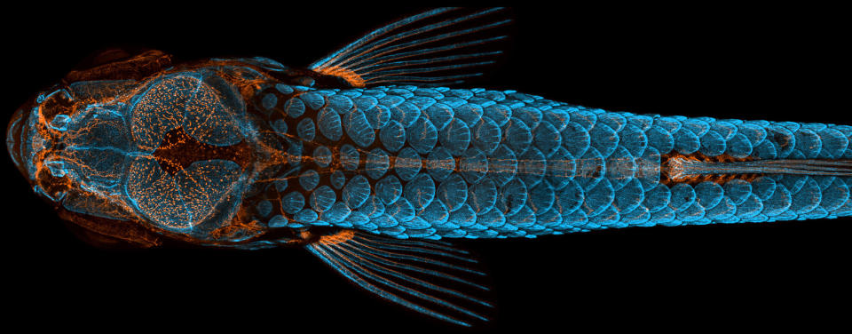
1st Place: Dorsal view of bones and scales (blue) and lymphatic vessels (orange) in a juvenile zebrafish.
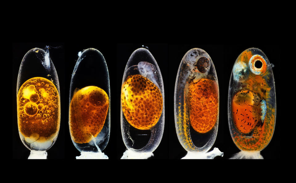
2nd Place: Embryonic development of a clownfish.
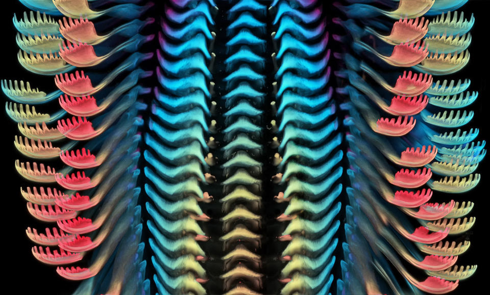
3rd Place: Tongue of a freshwater snail.
Love HuffPost? Become a founding member of HuffPost Plus today.
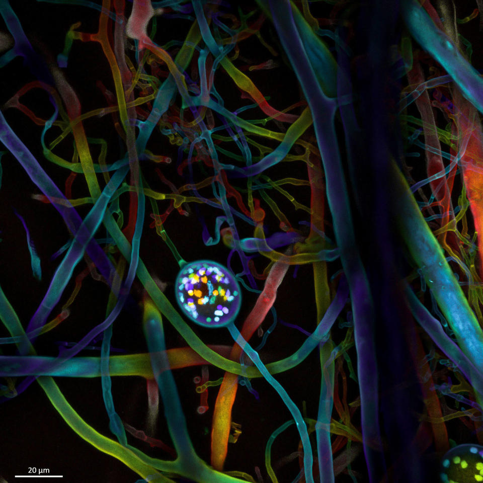
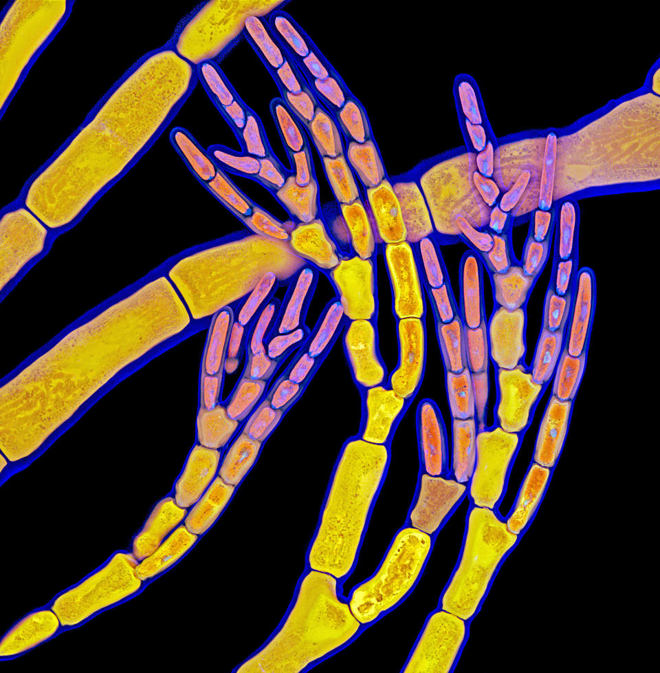
Multi-nucleate spores and hyphae of a soil fungus.
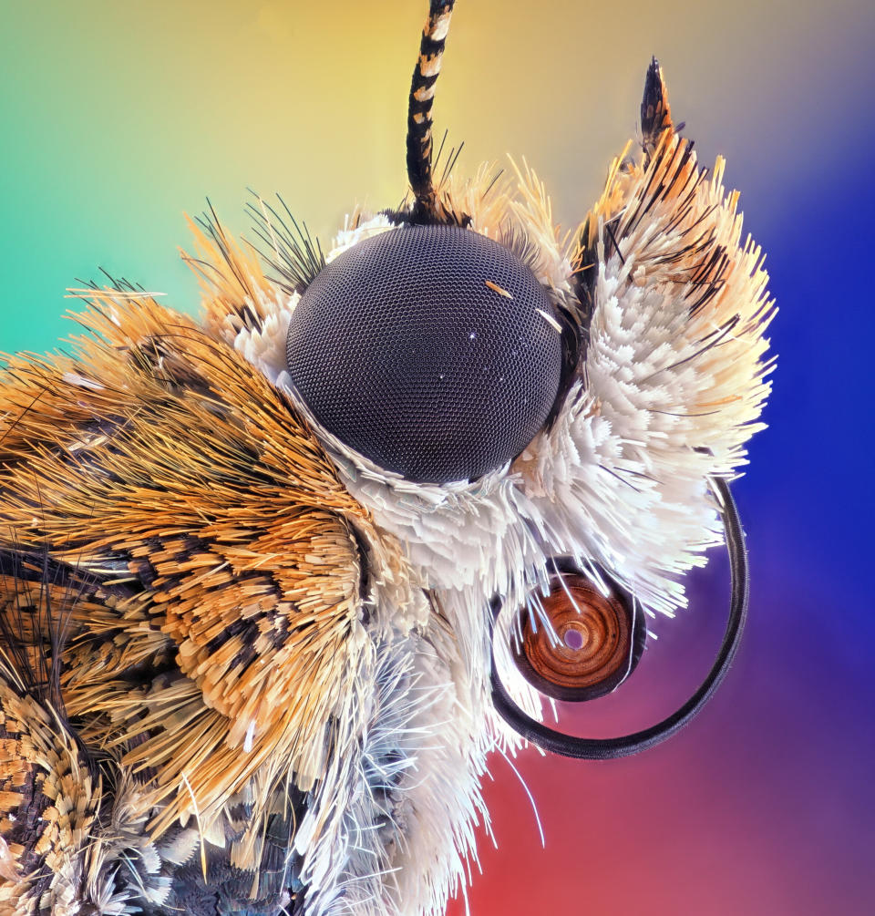
Bogong moth.
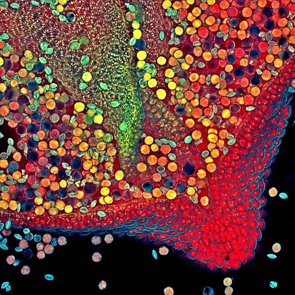
Hebe plant anther with pollen.
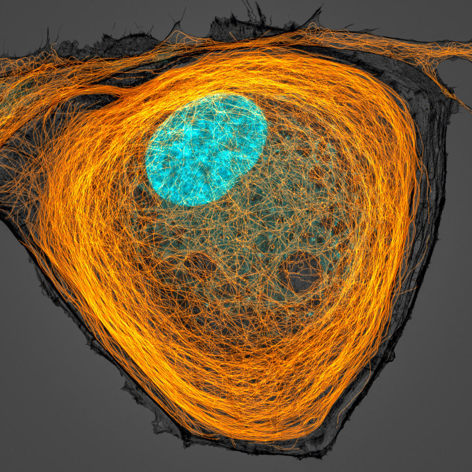
Microtubules (orange) inside a cell. Nucleus is shown in cyan.
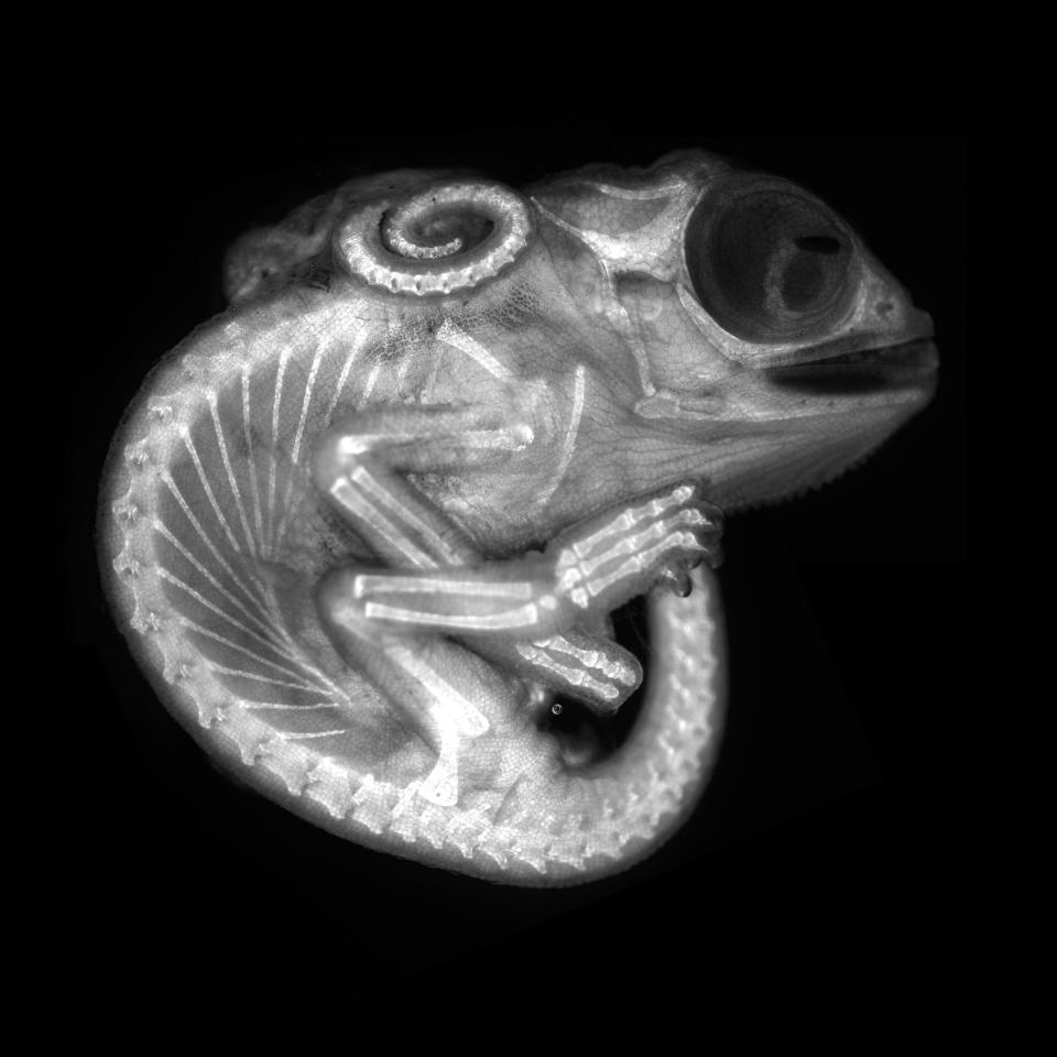
Chameleon embryo.

Connections between hippocampal neurons (brain cells).
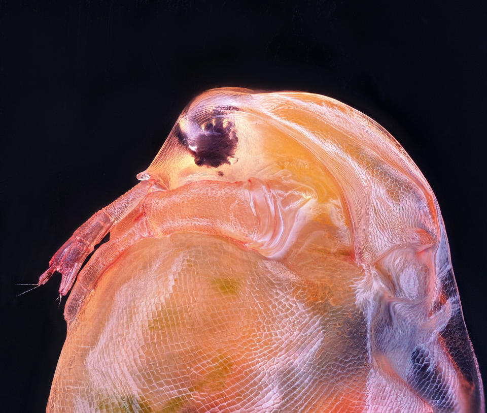
Daphnia magna, a small crustacean.
Red algae.
Human hair.

Crystals formed after heating an ethanol and water solution containing L-glutamine and beta-alanine.
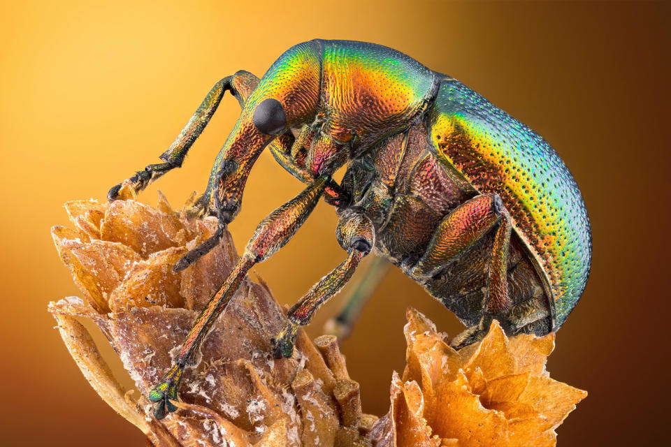
Leaf roller weevil.
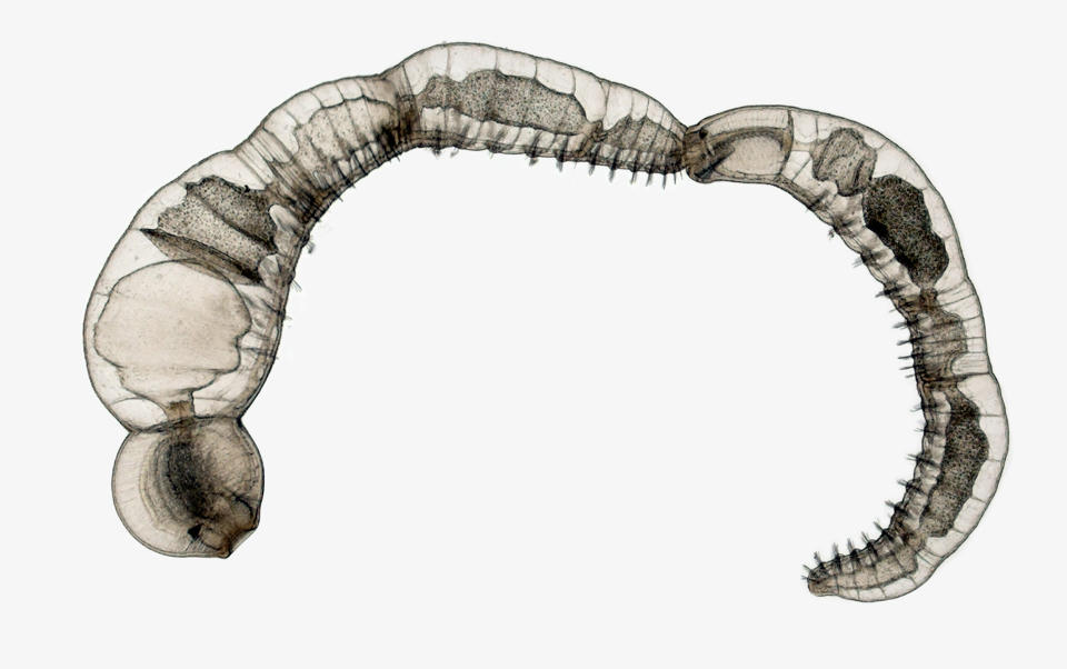
Chain of daughter individuals from the asexually reproducing annelid species.
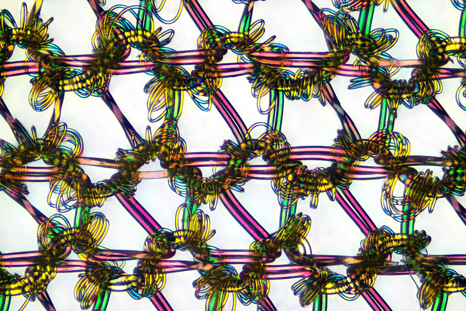
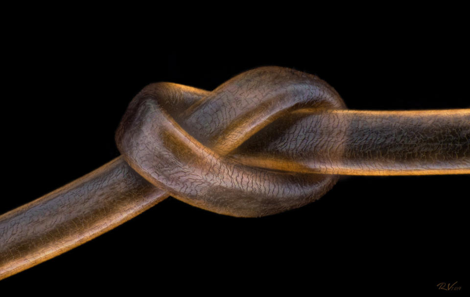
Nylon stockings.
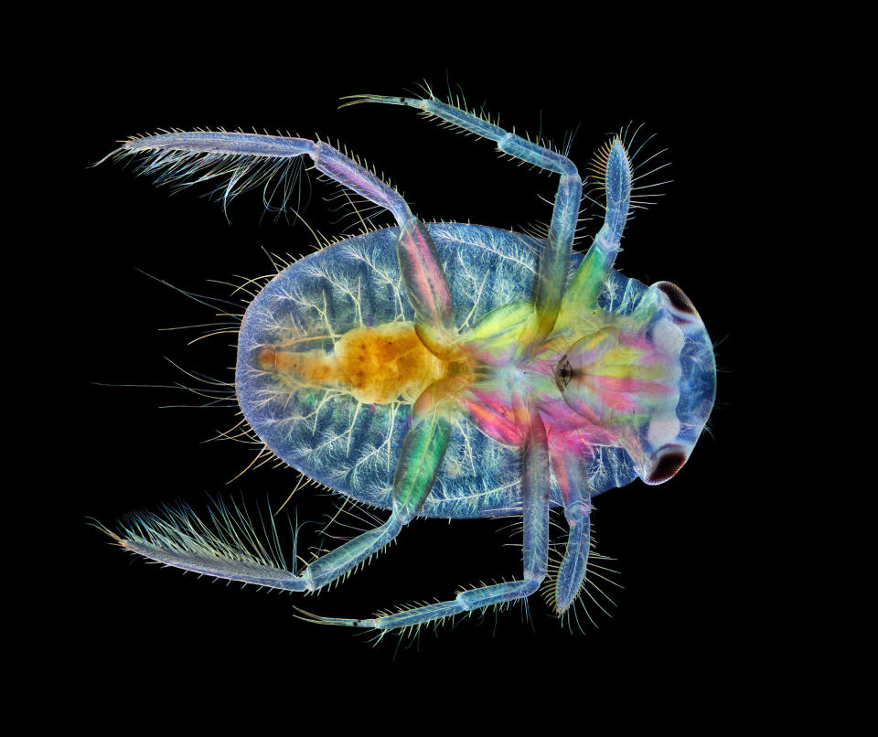
Ventral view of an immature water boatman.
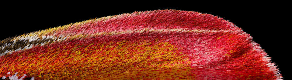
Atlas moth wing.
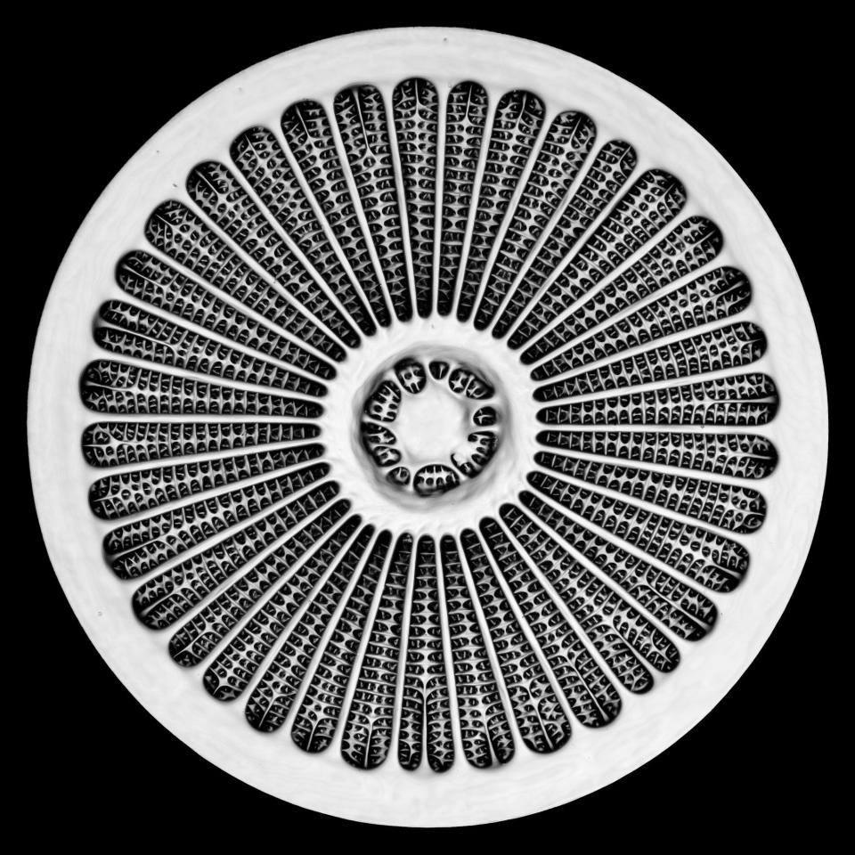
Silica cell wall of the marine diatom Arachnoidiscus sp.
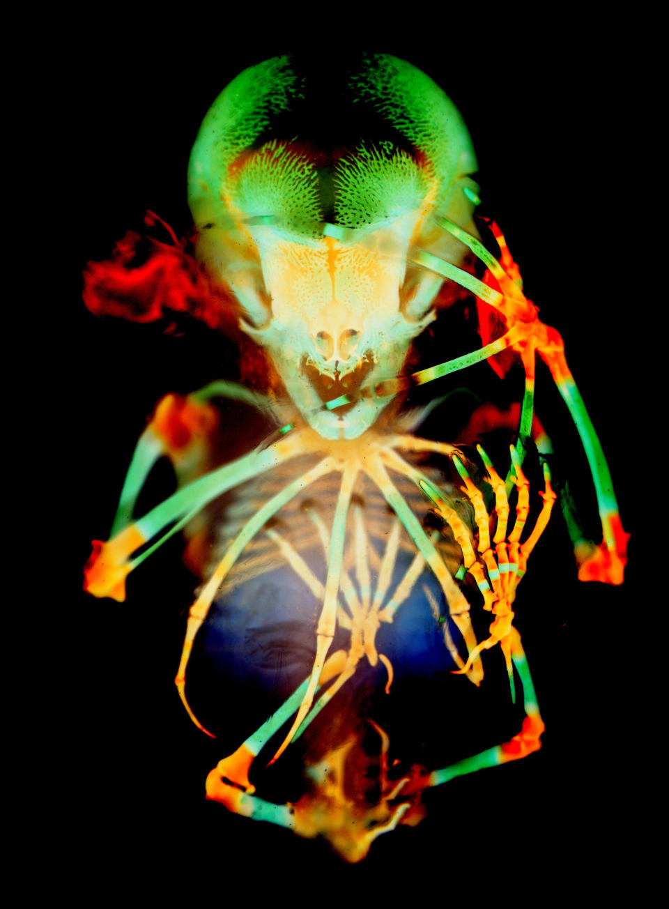
Skeleton preparation of a short-tailed fruit bat embryo.
This article originally appeared on HuffPost and has been updated.

