Controversial Fossil Claim Finds New Scientific Support
By Kate Wong
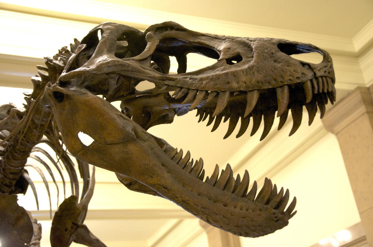
RALEIGH—Twenty years ago, paleontologist Mary Schweitzer made an astonishing discovery. Peering through a microscope at a slice of dinosaur bone, she spotted what looked for all the world like red blood cells. It seemed utterly impossible—organic remains were not supposed to survive the fossilization process—but test after test indicated that the spherical structures were indeed red blood cells from a 67-million-year-old Tyrannosaurus rex. In the years that followed, she and her colleagues discovered other apparent soft tissues, including what seem to be blood vessels and feather fibers. But controversy accompanied their claims. Skeptics argued that the alleged organic tissues were instead biofilm—slime formed by microbes that invaded the fossilized bone.
Schweitzer and her colleagues have continued to amass support for their interpretation. The latest evidence comes from a molecular analysis of what look to be bone cells, or osteocytes, from T. rex and Brachylophosaurus canadensis. The researchers isolated the possible osteocytes and subjected them to several tests. When they exposed the cell-like structures to an antibody that targets a protein called PHEX found only in bird osteocytes (birds are descended from dinosaurs), the structures reacted, as would be expected of dinosaur osteocytes. And when the team subjected the supposed dinosaur cells to other antibodies that target DNA, the antibodies bound to material in small, specific regions inside the apparent cell membrane.
Furthermore, using a technique called mass spectrometry, the investigators found amino acid sequences of proteins in extracts of the dinosaur bone that matched sequences from proteins called actin, tubulin and histone4 that are present in the cells of all animals. Although some microbes have proteins that are similar to actin and tubulin, the researchers note that soil-derived E. coli as well as sediments that surrounded the two dinosaur specimens failed to bind to the actin and tubulin antibodies that bound to the extract containing the apparent osteocytes.
Love HuffPost? Become a founding member of HuffPost Plus today.
Schweitzer and her collaborators detailed their findings in a paper released online October 16 in the journal Bone and in a talk given October 17 in Raleigh at the annual meeting of the Society of Vertebrate Paleontology. “Here’s the data in support of a biofilm origin,” Schweitzer said in her presentation as she showed a blank slide. “We haven’t found any yet.”
Skin And Bones
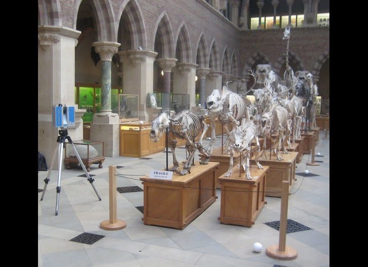
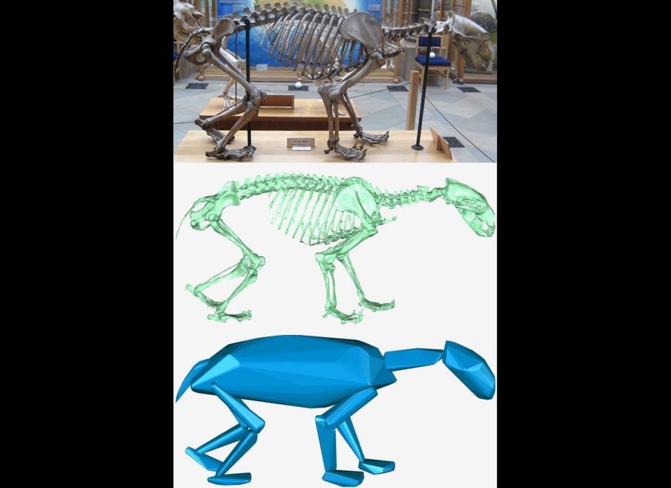
Scanning The Skeletons
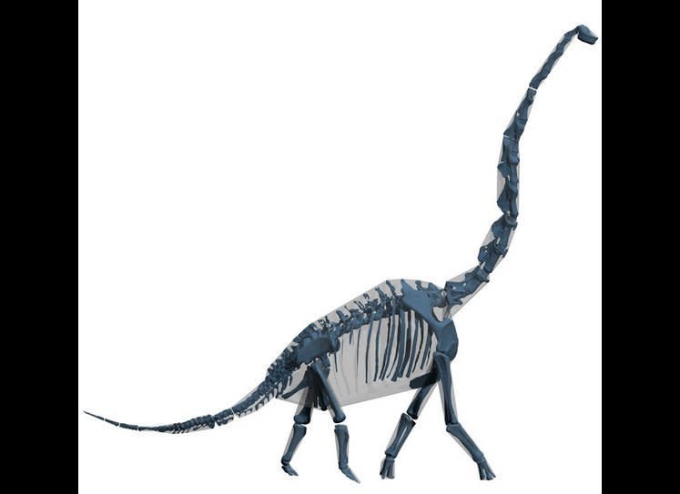
The Making Of A Digital Dino
3-D Dino
This article originally appeared on HuffPost.
